Micro-fluidic Raman chip and method for detecting exosome in blood based on micro-fluidic Raman chip

An exosome and microfluidic technology, applied in Raman scattering, chemical instruments and methods, biological testing, etc., can solve the problems of low stability and repeatability of immunoassay

Pending Publication Date: 2021-01-26
NINGBO UNIV
View PDF11 Cites 3 Cited by
  • Summary
  • Abstract
  • Description
  • Claims
  • Application Information

AI Technical Summary

Problems solved by technology

However, the stability and reproducibility of enhanced Raman-based immunoassays are low, and biomolecules, such as lipids, proteins, and nucleic acids are Raman-active and can exhibit stro

Method used


Image

  • Micro-fluidic Raman chip and method for detecting exosome in blood based on micro-fluidic Raman chip
  • Micro-fluidic Raman chip and method for detecting exosome in blood based on micro-fluidic Raman chip
  • Micro-fluidic Raman chip and method for detecting exosome in blood based on micro-fluidic Raman chip

Examples


Embodiment 1

[0057] A method for detecting exosomes in blood based on a microfluidic Raman chip. The method comprises the following steps:

[0058] 1. Preparation of exosome standard samples

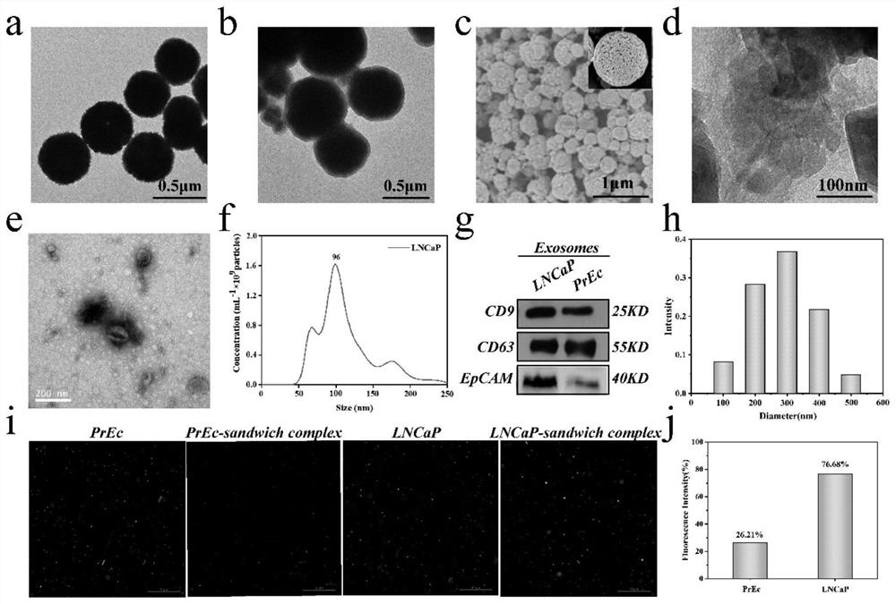
[0059] Cell culture: LNCaP and normal prostate epithelial cells PrEC were cultured at 37°C in a medium containing RPMI 1640 (10% (v / v) fetal bovine serum and 1% (v / v) penicillin-streptomycin). When the growth reached 100%, the medium was removed, the cells were washed with PBS (2X), and the medium was collected after 72 hours of serum-starved culture (RPMI 1640, without fetal bovine serum).

[0060] Exosome extraction: Collect cell culture fluid, centrifuge at 4°C (3000g, 20min) to remove cells and cell debris, then filter with a 0.22μm filter to completely remove large cell debris, then centrifuge at 4°C, 120,000g for 60min , and the supernatant was removed by centrifugation twice. Finally, 100 μL of PBS was added to obtain the prepared exosome solution, which was stored at −20 °C for further anal...

Embodiment 2

[0070] Embodiment 2 Optimization of chip channel and flow rate

[0071] In order to optimize the micro-column spacing and flow rate in the mixing area, four spacings of 150 μm, 200 μm, 250 μm, and 300 μm were designed to study their impact on capture efficiency. Exosomes labeled with PKH26 staining flowed through the chip, and the fluorescence intensities of inflow and outflow were calculated. To calculate the capture efficiency of exosomes. Such as Figure 4 As shown in c, the capture efficiency of exosomes decreases with the increase of the spacing, to prevent the clogging of the microchannel caused by the aggregation of magnetic beads, we use 200 μm as the optimal spacing of the microcolumns. 4d is the flow rate optimization diagram, using 6 different flow rates for research, in order to quickly detect the target, choose 0.6μL min -1 For the optimal flow rate, the capture efficiency of exosomes was as high as 72.5% within 1 h.

[0072] designed as Figure 7 chip shown. ...

Embodiment 4

[0079] Example 4 Detection of Exosomes in Blood Samples

[0080] Serum samples were collected from 8 prostate cancer patients and 10 normal subjects. This experiment was approved by the Ethics Committee of Shanghai Ninth People's Hospital, and written informed consent was obtained at the beginning of the project. The details are shown in Table 1.

[0081] Table 1. Information on clinical serum samples.

[0082] Patient ID TNM sex Age, years PLT, 10 9 / L

[0083] Serum samples were filtered through a 0.22 μm filter, and then detected using the chip and method prepared in Example 1. Raman spectra were obtained by detecting a 20 μL sample accurately injected at a constant flow rate by a micro flow syringe pump. According to the previous detection of exosome samples, the content of exosomes in the actual blood sample was calculated, such as Figure 6 As shown in a, the exosome content values ​​in the blood samples of prostate patients and normal people were...


PUM


Abstract

The invention discloses a micro-fluidic Raman chip and a method for detecting exosome in blood based on the micro-fluidic Raman chip, and belongs to the technical field of microfluidics. The micro-fluidic Raman chip is composed of four inlets, an outlet, a valve, a mixing chamber and a Raman detection area. Serum samples and CD63 antibody magnetic beads are pumped into the chip through an inlet 1and an inlet 3 respectively, and when two solutions flow through the staggered triangular micro-column mixing chambers, the two solutions are fully mixed and subjected to immunoreaction. And the CD63magnetic bead exosome compound is fixed in the Raman detection area through magnetic force. And the EpCAM functionalized Raman spheres are added from the inlet 4 to be mixed and reacted with the CD63magnetic bead exosome compound, and finall Raman detection is performed. The chip can be used for rapidly, simply, sensitively and quantitatively detecting the exosome in a serum sample, has strong detection specificity, is simple to operate, and is beneficial to more accurately evaluating the diagnosis of various diseases and guiding the treatment.

Description

technical field [0001] The array microfluidic chip designed in this study is used for the detection of exosomes in serum samples, and the chip can be used in the field of clinical cancer detection and analysis technology. Background technique [0002] Prostate cancer (PCa) is one of the most common cancers in men worldwide. PSA is the most widely used biomarker in prostate cancer screening and diagnosis. However, PSA is not a unique marker for prostate cancer. PSA levels also occur in non-cancerous diseases such as benign prostatic hyperplasia (BPH) and prostatitis. raised. Therefore, specific biomarkers are urgently needed to improve the specificity and sensitivity of PCa diagnosis. Exosomes are 30-200nm in diameter, are released by most cells, and exist in different biological fluids, such as blood, saliva, and urine. Exosomes reflect the specific physiological conditions and functions of their parent cells. Tumor-derived exosomes due to their high content (10 11 mL ...

Claims


Application Information

Patent Timeline
IPC IPC(8): G01N33/68G01N33/574G01N33/543G01N21/65B01L3/00
CPCG01N33/6872G01N33/57488G01N33/57434G01N33/56966G01N33/54326G01N21/65B01L3/50273G01N2333/70596
Inventor 王妍蔺施海梅田慧余绍宁
Owner NINGBO UNIV
Why Eureka
Try Eureka