Microscopic anastomosis training model and vascular model preparation process for model

A blood vessel model and training model technology, applied in the field of medical teaching aids, can solve the problems of poor simulation degree, complicated implementation process and high training cost, and achieve the effect of improving accuracy

Pending Publication Date: 2021-02-02
宁波创导三维医疗科技有限公司 +1
View PDF0 Cites 3 Cited by
  • Summary
  • Abstract
  • Description
  • Claims
  • Application Information

AI Technical Summary

Problems solved by technology

[0003] The current microvascular anastomosis training mainly focuses on suturing small rubber sheets and silicone tubes in the early stage, which is poor in simulation; in the later stage, anim

Method used


Image

  • Microscopic anastomosis training model and vascular model preparation process for model
  • Microscopic anastomosis training model and vascular model preparation process for model
  • Microscopic anastomosis training model and vascular model preparation process for model

Examples


Embodiment 1

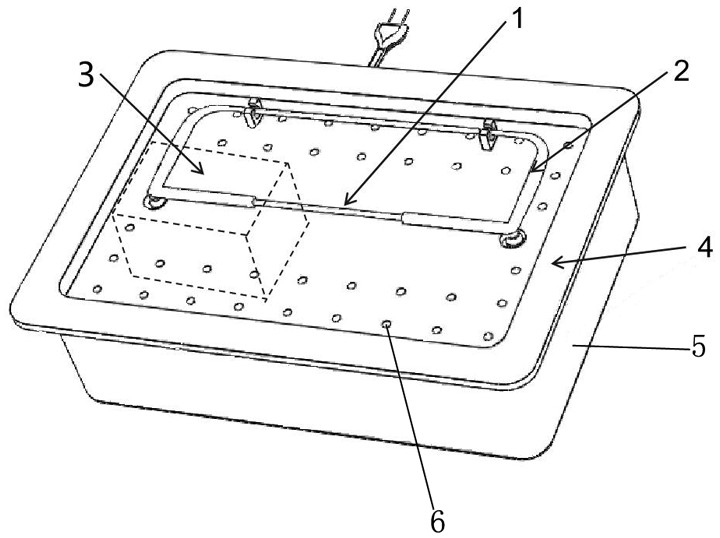
[0034] In this embodiment, the microanastomosis operation training model consists of, as figure 1 It includes a blood vessel model 1, a pipeline connection system 2, a blood circulation pump 3, a base 5 and a support platform 4; the support platform 4 is horizontally arranged on the upper end of the base 5; a sealed cavity is formed between the support platform 4 and the lower wall of the base 5 body; the blood circulation pump 3 is set in the aforementioned sealed cavity; the support platform 4 is provided with a number of openings 6 with a diameter of 2 mm; the pipeline connection system 2 and the blood vessel model 1 are set on the aforementioned support platform 4; the pipeline connection system 2 The fixation is realized by several buckles arranged on the support platform 4 ; the blood vessel model 1 is connected with the pipeline connection system 2 through joints; the pipeline connection system 2 is connected with the blood circulation pump 3 . The diameter of the pipel...

Embodiment 2

[0041] The composition of the microanastomosis training model in this embodiment is the same as that in Embodiment 1.

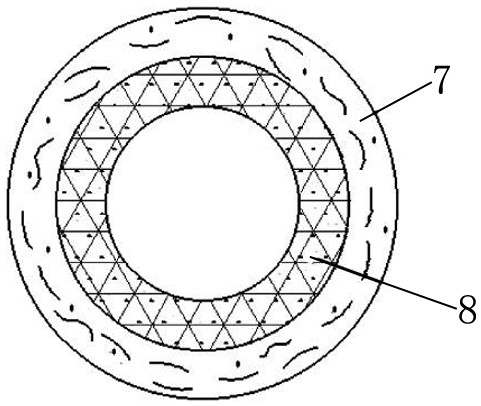
[0042] The vascular model 1 is a rough intimal small blood vessel classification such as image 3 As shown, from the outside to the inside, there are outer blood vessels and inner blood vessels. The inner diameter of the vessel is 0.8mm, and the total wall thickness from the inner vessel to the outer vessel is 1.5mm. The outer vessel layer 7 is smooth and complete, and the inner vessel vessel layer 8 is in the shape of rough plaques 9 under the microscope, and the plaques 9 are randomly distributed. The inner layer material is a mixture of Type I composite hydrogel and micron-sized calcified particles, simulating the texture and performance of vascular calcification in the early stage. The hardness of the material is Shore 0 00, the tensile strength is 2.5 MPa, and the elastic modulus is 4 MPa. Type II composite hydrogel is used for the outer layer 7 of the ...

Embodiment 3

[0048] In this embodiment, the composition of the training model for microanastomosis is the same as in Embodiment 1

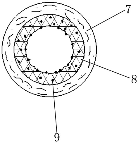
[0049] The vascular model 1 is the classification of small blood vessels with intimal separation as follows: Figure 4 , from the outside to the inside are the outer blood vessel, the sacrificial material layer and the inner blood vessel. The inner diameter of the vessel is 1.5mm, and the total wall thickness from the inner vessel to the outer vessel is 2mm. A separation section 10 is arranged between the inner layer and the outer layer of the blood vessel along the axial direction of the blood vessel to separate the inner layer of the blood vessel wall from the outer layer of the blood vessel wall. Finally, the separation of the inner and outer vessels is achieved. The blood vessel wall is smooth and complete, and the performance of the inner and outer layers is the same as that of normal small blood vessels. The inner layer 8 of the blood vessel is printe...


PUM

PropertyMeasurementUnit
The inside diameter ofaaaaaaaaaa
Tensile strengthaaaaaaaaaa
Elastic modulusaaaaaaaaaa

Abstract

The invention discloses a microscopic anastomosis training model and a vascular model preparation process for the model. The training model comprises a vascular model, a pipeline connecting system, ablood circulating pump, a base and a supporting platform; the blood circulating pump is arranged in a sealed cavity; a plurality of openings are formed in the supporting platform; the vascular model is communicated with the pipeline connecting system through a joint; and the pipeline connecting system is communicated with the blood circulating pump. The vascular model adopts a specific material and a specific manufacturing process, so that the touch feeling and the structure are close to those of real blood vessels of a human body; the microscopic anastomosis operation training model is helpful for better understanding blood vessel anatomy and anastomosis angles, directions, positions and effects, and the model is used for assisting a trainee to improve the accuracy of blood vessel anatomyseparation and suture. Moreover, after the model is displayed to the patient, the patient can visually and deeply understand physiology, anatomy, characteristics, operation schemes and the like of the neurosurgery operation.

Description

technical field [0001] The invention belongs to the field of medical teaching aids, and in particular relates to a microanastomosis operation training model and a preparation process of a blood vessel model for the model. Background technique [0002] Microsurgery is a surgical technique that uses special and fine instruments and materials to repair and reconstruct small blood vessels, nerves and other tissues under an operating microscope. It is characterized by small trauma, high surgical quality, and expanded surgical scope. It enables operations that cannot be performed directly under the naked eye, the most common of which is microvascular anastomosis. Microvascular anastomosis is an essential skill for doctors in trauma surgery, hand surgery, maxillofacial and plastic surgery, reconstructive surgery, etc. It is necessary for the survival of replanted fingers (limbs), thumb reconstruction and free flap transplantation condition. However, in order to master the techniq...

Claims


Application Information

Patent Timeline
IPC IPC(8): G09B23/30G09B9/00B29C64/118B29C64/20B29C64/209B29C64/241B29C64/314B29C64/336B29C64/40B29C64/393B33Y10/00B33Y30/00B33Y40/00B33Y40/10B33Y50/02
CPCG09B23/303G09B9/00B29C64/118B29C64/20B29C64/209B29C64/241B29C64/314B29C64/336B29C64/40B29C64/393B33Y10/00B33Y30/00B33Y40/00B33Y40/10B33Y50/02
Inventor 田磊杨勇张静宫海波毛茅徐方远
Owner 宁波创导三维医疗科技有限公司
Why Eureka
Try Eureka