A 3D-printed mechanical bionic auricular cartilage tissue engineering scaffold and its manufacturing method

A 3D printing, cartilage tissue technology, used in tissue regeneration, medical science, additive processing, etc., to save printing materials, reduce support structures, and have excellent printability.

Active Publication Date: 2022-07-26
INST OF BIOMEDICAL ENG CHINESE ACAD OF MEDICAL SCI
View PDF5 Cites 0 Cited by
  • Summary
  • Abstract
  • Description
  • Claims
  • Application Information

AI Technical Summary

Problems solved by technology

[0005] Although personalized scaffolds with fine structures can be obtained by 3D printing technology and used for the construction of tissue-engineered auricular cartilage, a problem that cannot be ignored is that the formation of cartilage tissue is affected by the mechanical properties of the scaffold. The ideal auricular cartilage tissue The engineered scaffold should have mechanical properties similar to those of the natural ear, give seed cells suitable biomechanical signals, and effectively maintain the shape and structural integrity of the engineered auricle tissue

Method used


Image

  • A 3D-printed mechanical bionic auricular cartilage tissue engineering scaffold and its manufacturing method
  • A 3D-printed mechanical bionic auricular cartilage tissue engineering scaffold and its manufacturing method
  • A 3D-printed mechanical bionic auricular cartilage tissue engineering scaffold and its manufacturing method

Examples


Embodiment 1

[0034] 1. Use 123D Design 2.2.14 to design a cube model of 8mm×8mm×3mm, choose a PLCL-PCL blend with a ratio of 90:10 as the raw material (CN201910139657.9), and print it by melt extrusion deposition (FDM). method, using a desktop 3D printer to print at 170°C, printing speed 2mm / s, and 70% filling conditions to obtain a cube model with a porous frameless structure, measure and print to obtain the actual three-dimensional size of the model, and according to the original model size data, The shrinkage rates on the X, Y and Z axes in 3D printing were calculated respectively, and the results were: length (X axis), 6.75% ± 0.51%; width (Y axis), 10.82% ± 0.43%; height (Z axis) ), 2.35%±0.73%.

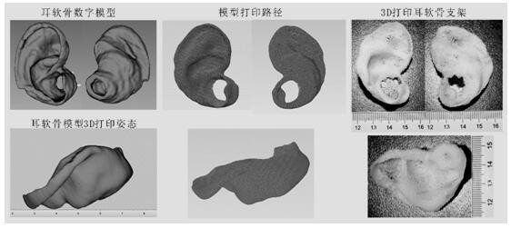
[0035] 2. In Materialise Magics 24.0, the acquired auricle cartilage scan file is converted into a digital model file and its contact angle with the horizontal plane is adjusted to maximize the contact area with the horizontal plane; according to the X, Y and Z calculated in step 1) The shr...

Embodiment 2

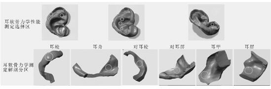
[0037] 1. In Magics 24.0, the digital model of the auricle cartilage adjusted in step 2) of Example 1 is divided into the digital model regions of the tragus, the antitragus, the helix, the antihelix, the concha and the auricular nave according to the anatomical structure. The maximum area of ​​the cylindrical area with a flat structure that can be selected is used as a reference, and the digital model areas of the tragus, the antitragus, the helix, the antihelix, the concha and the nave are selected to meet the mechanical performance test requirements of the stent. . Based on the shrinkage results of the 90:10 PLCL-PCL blend 3D printing 70% filling rate cube scaffolds in the X, Y and Z axes, the digital model of the selected area was scaled and adjusted according to the above process and conditions 3D Print borderless porous scaffolds.

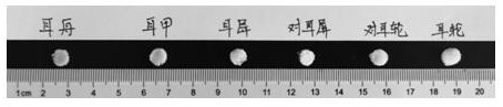
[0038] 2. The mechanical properties of the model bracket in the selected area of ​​the mechanical test obtained by 3D printing in step 1) a...

Embodiment 3

[0040] 1. Use 123D Design 2.2.14 to design a cube model with a porous frameless structure of 8mm × 8mm × 3mm, select a PLCL-PCL blend with a ratio of 80:20 as the raw material, and print it by melt extrusion deposition (FDM). , use a desktop 3D printer to print the model at a filling rate of 70% at 170 ° C and a printing speed of 2 mm / s, measure and print the actual three-dimensional size of the model, and calculate the X in 3D printing according to the size data of the original model. The shrinkage rate on the three axes of , Y and Z, the results are: length (X axis), 6.75% ± 0.51%; width (Y axis), 10.82% ± 0.43%; height (Z axis), 2.35% ± 0.73% .

[0041] 2. In Magics24.0, the acquired auricle cartilage scan file is converted into a digital model file and its contact angle with the horizontal plane is adjusted to maximize the contact area with the horizontal plane; according to the X, Y and Z three calculated in step 1) The shrinkage rate results on the axis, after scaling a...


PUM

PropertyMeasurementUnit
polydispersity indexaaaaaaaaaa

Abstract

The invention relates to a 3D printed mechanical bionic auricular cartilage tissue engineering scaffold and a manufacturing method thereof. It uses 3D printing technology to print a personalized auricular cartilage scaffold based on the patient's individual auricular cartilage structure model. Lactone (PLCL) and polycaprolactone (PCL) biodegradable composite material (PLCL-PCL) is printed, the auricular cartilage tissue engineering scaffold without borders, porous and through-holes of the present invention is biocompatible and degradable It has good stability and thermal stability, has a fine and complex three-dimensional structure of auricular cartilage, and has similar mechanical properties to the corresponding anatomical regions of natural human auricular cartilage tissue. It is suitable for the mechanical properties of natural auricular cartilage and can be applied Construction of personalized tissue-engineered auricular cartilage.

Description

technical field [0001] The invention relates to a 3D-printed mechanical bionic auricular cartilage tissue engineering support and a manufacturing method thereof, belonging to the technical field of tissue engineering. Background technique [0002] There is currently no satisfactory clinical treatment for ear cartilage defects caused by congenital malformations, trauma, infection, tumors, and surgery. Tissue engineering technology provides a promising approach for the repair and reconstruction of auricular cartilage. Generally, tissue engineering technology is to plant seed cells on tissue engineering scaffolds to construct a three-dimensional complex of cells and biomaterials. Among them, the tissue engineering scaffold is one of the three elements of tissue engineering construction. The ideal tissue engineering scaffold should have good biocompatibility and biodegradability, a three-dimensional porous structure with suitable volume and shape, high porosity and internal po...

Claims


Application Information

Patent Timeline
Patent Type & Authority Patents(China)
IPC IPC(8): A61L27/50A61L27/56A61L27/26B33Y10/00B33Y70/00B33Y80/00
CPCA61L27/50A61L27/56A61L27/26B33Y10/00B33Y70/00B33Y80/00A61L2430/14C08L67/04
Inventor 李学敏王艺蒙王自强张逸芸段瑞平杜博刘玲蓉
Owner INST OF BIOMEDICAL ENG CHINESE ACAD OF MEDICAL SCI
Why Eureka
Try Eureka