Thyroid nodule diagnosis method based on deep learning network

A deep learning network, thyroid nodule technology, applied in the field of image processing and artificial intelligence-assisted diagnosis of diseases, can solve problems such as difficulty in obtaining better results, high noise in ultrasound images, and lack of doctor resources, reducing time and work. Intensity and stress, the effect of reducing economic and psychological burden

Active Publication Date: 2021-03-19
XUZHOU MEDICAL UNIV
View PDF14 Cites 15 Cited by
  • Summary
  • Abstract
  • Description
  • Claims
  • Application Information

AI Technical Summary

Problems solved by technology

Ultrasound has become the first choice for diagnosis because of its high sensitivity, non-invasive, non-radiation, and real-time characteristics. For thyroid nodules that cannot be characterized by ultrasound, invasive FNA (fine-needle aspiration biopsy) should be used to confirm. However, the uncertainty rate of FNA It is also as high as 30%, and most doctors often choose conservative treatment for unnecessary surgical resection, causing patients to take drugs for life, causing irreparable harm to patients
[0004] According to statistics, the rate of misdiagnosis and missed diagnosis of thyroid nodules by ultrasound is as high as 34%. In addition, due to the huge outpatient volume of thyroid diseases, each ultrasound doctor needs to diagnose 120-150 patients per day on average, and the whole process of ultrasound diagnosis lasts for 20 minutes. It takes about 10 hours a day to focus on film reading, which is a huge workload
In addition, there is currently a shortage of 200,000 doctors specializing in ultrasound in the country, and the shortage of doctor resources
[0005] Due to the large amount of noise in ultrasound images and the significant differences in the size and location of thyroid glands in different populations, traditional image processing methods are often difficult to obtain better results.

Method used


Image

  • Thyroid nodule diagnosis method based on deep learning network
  • Thyroid nodule diagnosis method based on deep learning network
  • Thyroid nodule diagnosis method based on deep learning network

Examples


Embodiment 1

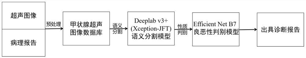
[0051] Such as figure 1 As shown, an embodiment of a method for diagnosing a thyroid nodule based on a deep learning network of the present invention comprises the following steps:

[0052] A. Establishment of Thyroid Ultrasound Image Database

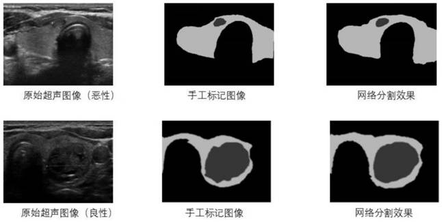
[0053] By collecting ultrasound images and pathology reports of thyroid patients in the pathology department and radiology department, the thyroid nodules were outlined and labeled using the labelme tool of Anaconda2 and the data was saved in josn format for subsequent use.

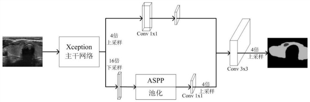
[0054] B Deeplab v3+ semantic segmentation based on Xception-JFT

[0055] a. Input the preprocessed ultrasound image into the segmentation model of Xception-JFT with Xception as the backbone network;

[0056] b. The network first performs feature extraction and downsampling through a 3×3 convolutional layer, and at the same time, the feature matrix is ​​input into the Decoder part after four times downsampling;

[0057] c. After the feature extraction is complete...

Embodiment 2

[0064] In other specific implementations of the present invention, the rest are the same as the above-mentioned implementations, the difference is that, as figure 1 As shown, the preprocessing process of the image marked in step A can enhance the stability of the network operation; the preprocessing includes the following steps

[0065] a. Process the marked image with python and opencv to generate two labeled images in BMP format, and set different pixel values ​​for thyroid nodules, parenchyma and other parts, so as to display different brightness to distinguish each part of the thyroid;

[0066] b. Generate a grayscale image from the marked image. The value of the gray-white channel is the matrix obtained by multiplying the grayscale image with the corresponding elements of the feature matrix of the mask of the thyroid parenchyma containing nodules. The gray channel only contains nodules. For multiple In the case of nodules, each nodule is distributed on the red channel of ...

Embodiment 3

[0070] In other specific implementations of the present invention, the rest are the same as the above-mentioned implementations, the difference is that, as Figure 4 As shown, Efficient Net B7 is selected for classification deep learning network:

[0071] a. Through the Efficient Net B7 model, the preprocessed input thyroid ultrasound image is subjected to feature extraction and reduction through a 3×3 convolution kernel,

[0072] b. Then use seven sets of moving reverse bottleneck convolutions to perform feature extraction at different scales,

[0073] c. Finally, use the fully connected layer to integrate features and use softmax to classify,

[0074] d. Output a two-dimensional vector containing benign probability and malignant probability of thyroid nodules to complete the judgment of benign and malignant thyroid nodules.

[0075] Efficient Net is a new model scaling method with extremely high parameter efficiency and speed. This method uses a simple but efficient compou...


PUM


Abstract

The invention discloses a thyroid nodule diagnosis method based on a deep learning network, and belongs to the field of image processing and artificial intelligence aided disease diagnosis. The methodcomprises the following steps: searching an ultrasonic original image and a pathological report of a thyroid nodule of a thyroid patient, and constructing a thyroid nodule database; preprocessing theultrasonic image; performing semantic segmentation on the ultrasonic image preprocessed in the step 2 through a Deeplab v3+ method based on Xception-JFT, and forming a semantic segmentation result graph; judging benign and malignant thyroid nodules based on a deep learning network; and forming a thyroid nodule diagnosis information report. According to the method, the Deeplab v3+ algorithm basedon Xeption-JFT is adopted to establish the thyroid ultrasound image segmentation network model, the optimal segmentation effect is achieved by continuously improving the backbone network Xception, nodule information can be automatically and rapidly recognized under high accuracy and high robustness, image features are automatically extracted for accurate segmentation to obtain a better diagnosis result, and an objective reference is provided for clinical diagnosis.

Description

technical field [0001] The invention belongs to the field of image processing and artificial intelligence-assisted diagnosis of diseases, and in particular relates to a method for diagnosing thyroid nodules based on a deep learning network. Background technique [0002] Thyroid nodules are lumps in the thyroid gland. The causes of the disease are complex and the number of patients is large. According to the survey, 68% of the patients in the country are potential patients with thyroid nodules. At the same time, the phenomenon of overdiagnosis and treatment is serious, and it has gradually become one of the diseases that threaten human health. [0003] Currently, nodules can be diagnosed by physician palpation, needle biopsy, and ultrasound images. Ultrasound has become the first choice for diagnosis because of its high sensitivity, non-invasive, non-radiation, and real-time characteristics. For thyroid nodules that cannot be characterized by ultrasound, invasive FNA (fine-n...

Claims


Application Information

Patent Timeline
Patent Type & Authority Applications(China)
IPC IPC(8): G06T7/00G06T7/12G06T7/13G06F16/53G06N3/04G06N3/08
CPCG06T7/0012G06T7/12G06T7/13G06F16/53G06N3/08G06T2207/10132G06N3/045
Inventor 唐璐徐凯张珂赵英红
Owner XUZHOU MEDICAL UNIV
Why Eureka
Try Eureka