A class of anti-human EGFR antibody drug conjugates and its preparation method and application

A technology of antibody drugs and antibody-drug conjugates, applied in the direction of antibodies, drug combinations, anti-tumor drugs, etc., to achieve enhanced anti-tumor effect, considerable yield, stable and controllable quality

Active Publication Date: 2022-05-10
MEDICINE & BIOENG INST OF CHINESE ACAD OF MEDICAL SCI
View PDF6 Cites 0 Cited by
  • Summary
  • Abstract
  • Description
  • Claims
  • Application Information

AI Technical Summary

Problems solved by technology

[0006] (3) Self-decomposition part: aminobenzyloxycarbonyl (p-aminobenzyloxycarbonyl) spacer is similar to an ester bond, and its stability in plasma needs to be further improved

Method used


Image

  • A class of anti-human EGFR antibody drug conjugates and its preparation method and application
  • A class of anti-human EGFR antibody drug conjugates and its preparation method and application
  • A class of anti-human EGFR antibody drug conjugates and its preparation method and application

Examples


Embodiment 1

[0058] The synthesis of embodiment 1 Linker-MMAE

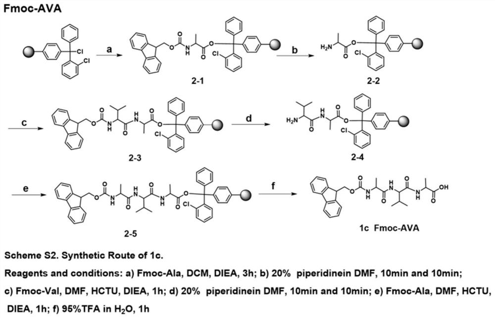
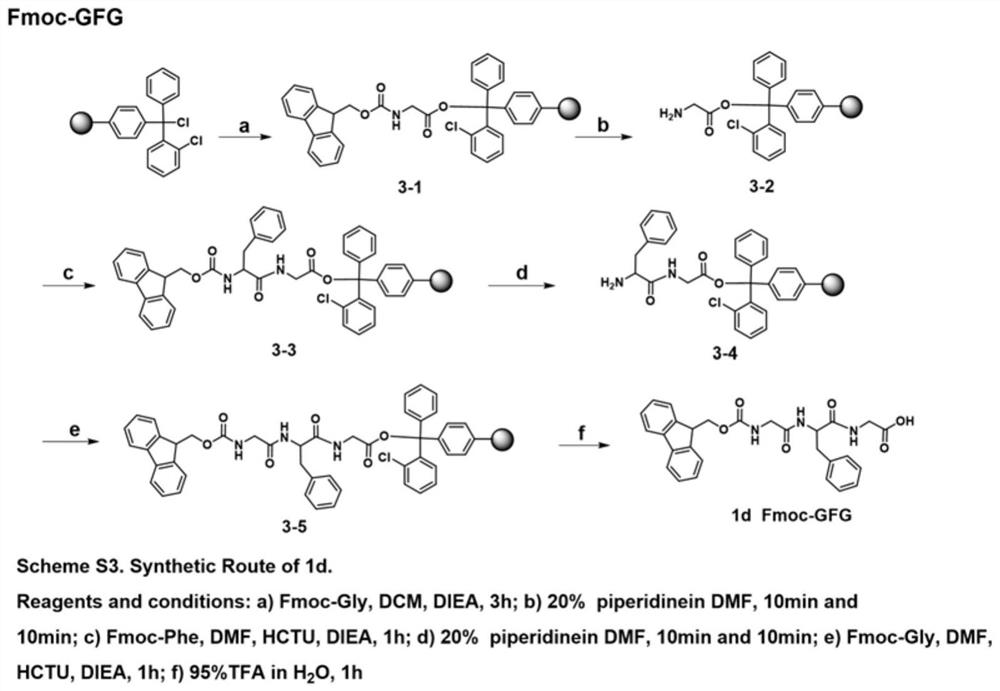
[0059] 1.1 Synthesis of Fmoc-AA

[0060] 1.1.1 Synthesis of Fmoc-dipeptide

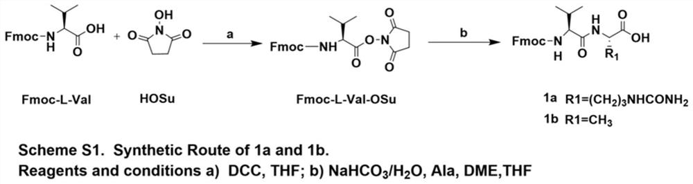
[0061] Synthesis of Fmoc-L-Val-OSu

[0062] according to figure 1 As shown in the Scheme S1 step, the reaction products Fmoc-L-Val (10g, 29.3mmol) and HoSu (3.7g, 32.3mmol) were dissolved in 100mL THF, and DCC (6.6g, 32.3mmol) was added under ice-bath conditions, After the addition was complete, the ice bath was removed, and the reaction was stirred overnight at room temperature. After the reaction was completed (petroleum ether: ethyl acetate = 1:1 detection), the reaction solution was moved to an ice bath, and after the solid was completely precipitated, it was filtered with suction, and the filtrate was rotary evaporated until it became foamy, and the residue was the product. The reaction product was directly used in the next reaction without further purification.

[0063] Synthesis of 1a

[0064] according to figure 1 As shown in Scheme S1,...

Embodiment 2

[0176] Example 2 In vitro plasma stability detection of Linker-MMAE

[0177] Linker-MMAE to be tested was prepared into a 10mM DMSO stock solution for use. 4 μL DMSO stock solution was added to 996 μL blank rat plasma, mixed evenly and placed in a 37°C water bath for incubation. 50 μL samples were taken at 0, 30, 60, 180, and 360 minutes respectively, and the reaction was terminated. Two parallel samples at each reaction time point were stored at -20°C and processed uniformly after the sampling was completed. The sample was centrifuged to settle protein, and the supernatant was taken, and the concentration of MMAE was detected by LC-MS / MS, and the concentration unit (nM) was detected. Draw the curve of the release amount of MMAE as a function of time, and calculate the release rate of MMAE in plasma. The release rate is shown in Table 1.

[0178] The results showed that spacer fraction M-2 was the best (K=4.0 nM / hr), followed by M-3 (K=10.9 nM / hr) and M-1 (K=22.6 nM / hr). Co...

Embodiment 3

[0181] Embodiment 3 Linker-MMAE digestion release rate experiment

[0182] Linker-MMAE to be tested was prepared into a 10mM DMSO stock solution for use. 10 μL of the stock solution was added to 2490 μL of 100 mM L-Cys PBS buffer (pH=6) to prepare a working buffer solution with a concentration of 0.04 mM. Cathepsin B, purchased at a concentration of 1.52 mM, was diluted with PBS buffer at pH=6 to form a 0.4 mM protease solution. 1000 μL of 0.04 mM compound buffer working solution was mixed with 10 μL of 0.4 mM protease solution (the molar ratio of substrate to protease was 1:10), and incubated in a 37°C water bath. The reaction was terminated at 0, 10, 60, 120, 240, and 360 minutes, respectively, and two parallel samples were taken at each reaction time point. The concentration of MMAE was detected by LC-MS / MS, and the detection concentration unit (nM).

[0183] The results are shown in Table 2. M-2 has the fastest hydrolysis release (K=32.3nM / hr), followed by C-4 (K=32.1nM...


PUM


Abstract

The invention discloses a kind of anti-human EGFR antibody drug conjugate, its preparation method and application. The anti-human EGFR antibody drug conjugate has the structure shown in formula I. The present invention uses SCT‑200 fully humanized monoclonal antibody to couple with Linker‑MMAE by reducing the disulfide bond between the antibody chains, and constructs a class of anti-human antibodies targeting EGFR for the treatment of solid tumors. EGFR Antibody Drug Conjugate. Compared with SCT-200 itself, the novel antibody-drug conjugate SCT-200-Linker-MMAE proposed by the present invention does not affect the affinity, endocytic activity and targeting of the antibody, and better retains its biological function. Compared with SCT-200, the activity has been significantly improved, and it can significantly inhibit the expression of related proteins in the process of tumor apoptosis, and the anti-tumor effect is greatly enhanced.

Description

technical field [0001] The invention relates to an anti-human epidermal growth factor receptor (EGFR) antibody-drug conjugate (ADC), including materials using the ADC, a preparation method and application in the field of solid tumor treatment. The invention belongs to the field of biotechnology medicine. Background technique [0002] EGFR (epidermal growth factor receptor) is a very important target in tumor therapy, and it is overexpressed in various solid tumors of epithelial origin. The main focus of anti-tumor therapy. Anti-EGFR antibody is the main targeted therapy, and its mechanism of action is mainly to compete with endogenous ligands for binding to EGFR, and to produce anti-tumor effects by inhibiting the activation of tyrosine kinase and promoting the internalization of EGFR. Although antibody drug research has its own unique advantages and good market prospects, the existing EGFR drugs have limited curative effects, and in most cases targeting EGFR, long-term us...

Claims


Application Information

Patent Timeline
Patent Type & Authority Patents(China)
IPC IPC(8): A61K47/68A61K38/08A61P35/00
CPCA61K47/6817A61K47/6873A61P35/00A61K38/08A61K47/68A61K39/395A61K47/55A61K47/65
Inventor 李卓荣胡馨月姜海伦刘睿白炜琪刘秀筠苗庆芳
Owner MEDICINE & BIOENG INST OF CHINESE ACAD OF MEDICAL SCI
Why Eureka
Try Eureka