

Atomic force microscope liquid phase imaging method
A technology of atomic force microscope and imaging method, which is applied in the direction of scanning probe microscopy, instruments, measuring devices, etc., can solve problems such as complicated operation, and achieve the effect of simple operation, effective operation and universal use
- Summary
- Abstract
- Description
- Claims
- Application Information
AI Technical Summary
Problems solved by technology
Method used
Image

Examples
Embodiment 1
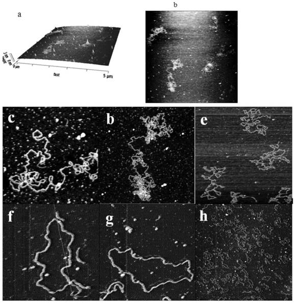
[0051] (1) Use epoxy glue to fix mica slices (circular and 1x1cm in diameter) on the glass slide at the bottom of the liquid pool 2 Square is applicable), let the epoxy resin cure after 12 hours;
[0052] 7U / mL of BspMI with λ-DNA in 10mM CaCl 2 , 25mM KCl, 10mM HEPES buffer, mixed evenly, reacted for 30 minutes, and the reaction temperature was 37°C;
[0053] Drop onto the newly dissociated mica sheet (the mica sheet is fixed on the bottom of the liquid pool or on the slide at the bottom of the BioCell), settle for 2 minutes, and then wash with 10mL of 10mMCaCl 2 , 25mMKCl, 10mM HEPES buffer for gentle washing, and finally wash with 2mL of 10mMNiCl2, 25mMKCl, 10mM HEPES imaging buffer, and add imaging buffer;
[0054] (2) There is no need to adjust the laser. After the sample is placed, use the needle directly in contact mode. The system will automatically stop when the gas-liquid interface is blocked. At this time, use z stepper Motors to manually place the needle 100 μm t...
PUM
 Abstract
Description
Claims
Application Information