Method for in-vitro detection of miRNA based on ratio fluorescence of acridine orange and carbon dots
A ratiometric fluorescence and in vitro detection technology, applied in the field of miRNA in vitro detection, can solve the problems of complex components, background fluorescence interference system, quenching, etc., and achieve the effect of good repeatability and broad application prospects
- Summary
- Abstract
- Description
- Claims
- Application Information
AI Technical Summary
Problems solved by technology
Method used
Image
Examples
Embodiment 1
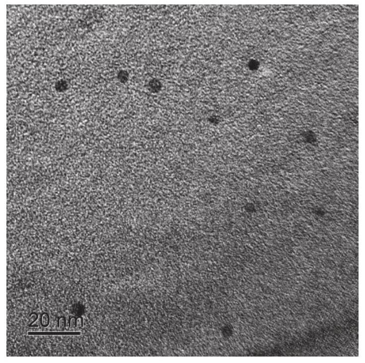
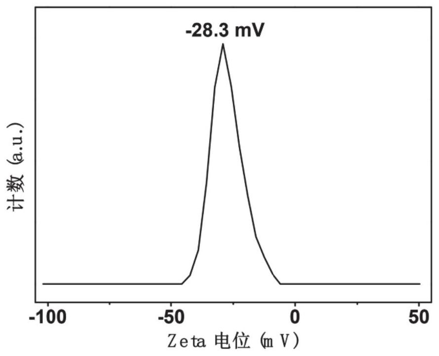
[0043] Preparation method of carbon dots: Dissolve 1.8 g of citric acid in 30 mL of formamide and transfer it to a stainless steel reaction kettle lined with polytetrafluoroethylene, react at 180 ° C for 6 hours, wait for the reaction kettle to naturally cool to room temperature and then add it to the reaction solution. Add 120 mL of acetone and put it in the refrigerator, place it at -20 °C for 24 hours to generate carbon dot precipitation, centrifuge at 10,000 rpm for 20 minutes to separate the carbon dot precipitation, and add the carbon dot precipitation to 30 mL of methanol / acetone with 10% methanol volume. In the mixed solution, centrifuge and wash at 10,000 rpm for 20 minutes, repeat the centrifugation and washing for 3 times, and then re-dissolve the carbon dots in 10 mL of methanol, and filter them with a filter membrane with a pore size of 0.22 μm to remove large particles. Add 30 mL of acetone, wash by centrifugation at 10,000 rpm for 20 minutes, and dry at 50° C. fo...
Embodiment 2
[0048] Add 3 μL of miRNA solution to 292 μL of DNA probe solution with a concentration of 10 nM to make the final concentration of miRNA to be 8.5 nM. After incubating at 37°C for 90 minutes, add 3 μL of acridine orange solution with a concentration of 1.76 μM and adsorb for 10 minutes. Then add 2 μL of carbon dot solution with a concentration of 0.1 mg / mL and let it stand for 10 minutes. Test the fluorescence of the solution with a fluorescence spectrophotometer. Figure 5 The standard curve equation in the calculated miRNA concentration was 8.43 nM, which was close to the actual concentration of miRNA. The detection results show that the detection method has high accuracy.
Embodiment 3
[0050] 3 μL of three control miRNAs (sequences from 5' to 3', No.1 (hsa-miR-223-3p): UGUCAGUUUGUCAAAUACCCCA, No.2 (miR- 92a-3p-TMT): UUUUCCACUUGUCCCGGCCUGU, No.3 (miR-92a-3p-SMT): UAUUCCACUUGUCCCGGCCUGU) solution to make the final concentration of the control miRNA to be 7nM, after incubation at 37°C for 90 minutes, 3 μL of 1.76 μM was added to it. The acridine orange solution was adsorbed for 10 minutes, then 2 μL of carbon dot solution with a concentration of 0.1 mg / mL was added and left for 10 minutes, and the fluorescence of the solution was tested with a fluorescence spectrophotometer. Figure 5 The standard curve equations in the calculated concentrations of the three control miRNAs were -0.12, -0.19, and 0.01 nM, which were close to 0. The detection results show that the detection method has good specificity.
PUM
| Property | Measurement | Unit |
|---|---|---|
| concentration | aaaaa | aaaaa |
| diameter | aaaaa | aaaaa |
Abstract
Description
Claims
Application Information
 - R&D
- Intellectual Property
- Life Sciences
- Materials
- Tech Scout
- Unparalleled Data Quality
- Higher Quality Content
- 60% Fewer Hallucinations
Browse by: Latest US Patents, China's latest patents, Technical Efficacy Thesaurus, Application Domain, Technology Topic, Popular Technical Reports.
© 2025 PatSnap. All rights reserved.Legal|Privacy policy|Modern Slavery Act Transparency Statement|Sitemap|About US| Contact US: help@patsnap.com


