Bionic micro-fluidic chip for simulating in-vivo microorganism-intestine-brain axis signal transduction process
A microfluidic chip, signal transduction technology, applied in the determination/inspection of microorganisms, the method of supporting/immobilizing microorganisms, the method of stress-stimulating the growth of microorganisms, etc.
- Summary
- Abstract
- Description
- Claims
- Application Information
AI Technical Summary
Problems solved by technology
Method used
Image
Examples
Embodiment 1
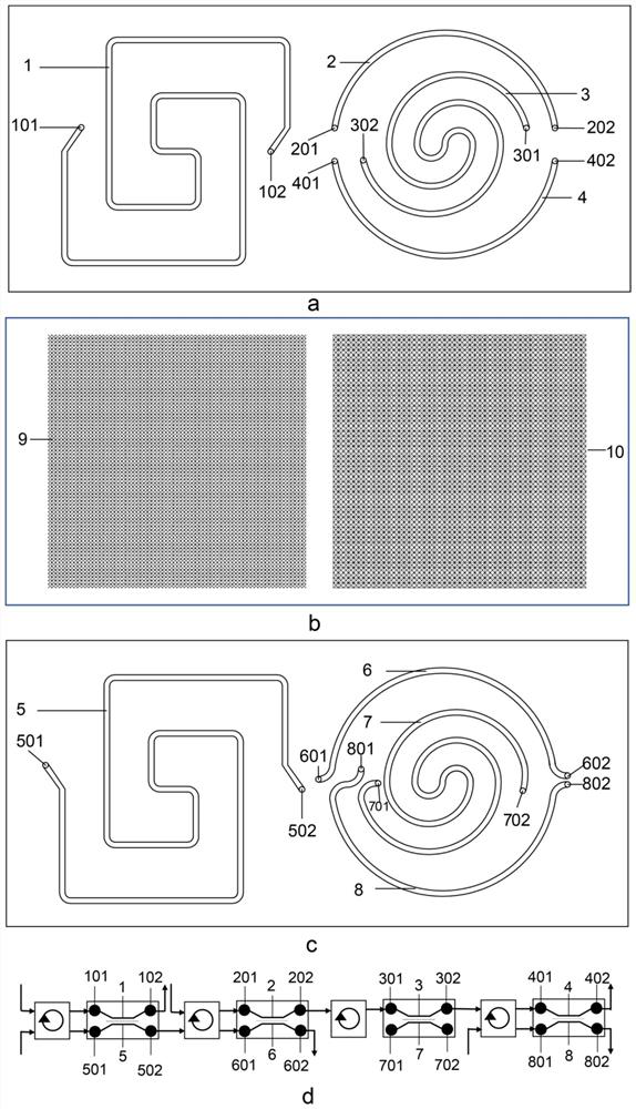
[0036] Design and fabricate microfluidic chips, such as figure 1 shown.
[0037] The microfluidic chip is mainly composed of a top chip, a nanoporous membrane, a microporous membrane and a bottom chip. The top chip is divided into left and right parts. The main passage entrance (101) and the first back-shaped main passage exit (102) are composed, and the right part of the top chip is composed of the first upper semicircular passage (2), the first upper semicircular passage entrance (201) and the first Upper semicircular channel outlet (202), the first helical main channel (3), the first helical main channel inlet (301) and the first helical main channel outlet (302), the first lower semicircular channel ( 4) and the first lower semicircular channel entrance (401) and the first lower semicircular channel outlet (402); the bottom chip is also divided into left and right parts, and the left part of the bottom chip is composed of the second back-shaped main channel ( 5), the sec...
Embodiment 2
[0047] Integrity Identification of MGB Axis Bionic Microfluidic Chip
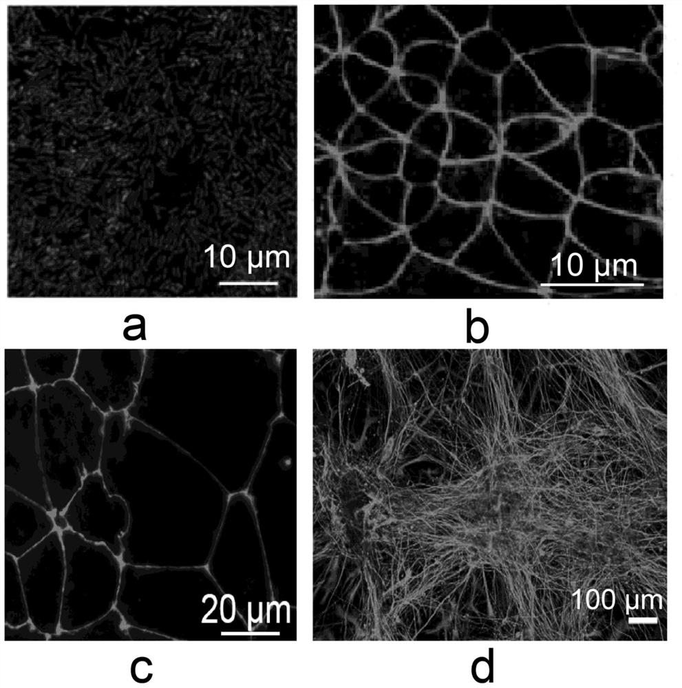
[0048] The two-layer PDMS of the microfluidic chip after 5 days of culture and operation was disassembled, and the intestinal epithelial cells Caco-2 and intestinal microorganisms co-cultured on the nanoporous membrane of the intestinal unit were stained for live dead cells and observed by fluorescence microscopy its coexistence state, such as figure 2 As shown in a, it can be seen that intestinal microorganisms and Caco-2 cells can coexist in the intestinal unit. Routine immunofluorescence staining was used to characterize the expression of ZO-1 protein on the surface of intestinal barrier Caco-2 cells and blood-brain barrier hBMECs cells, such as figure 2 As shown in b and 2c, it can be seen that tight junctions have been formed and have a barrier structure; the differentiation of human hippocampal neural stem cells in the bottom chip of the brain unit was observed by confocal fluorescence microscopy, ...
Embodiment 3
[0050] Permeability identification of MGB axis biomimetic microfluidic chip
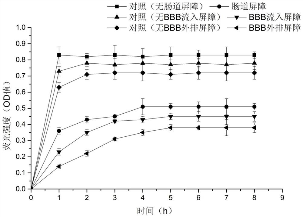
[0051] Add FITC-labeled dextran to the channel (1) of the top chip of the intestinal unit, and collect the bottom channel outlet (502) and The perfusate of the top channel outlet (202) of the BBB inflow unit and the top channel outlet (402) of the BBB efflux unit was detected in a microplate reader, and the absorption wavelength was 490nm. The blank control group was MGB without cell attachment Axis biomimetic microfluidic chip. Such as image 3 As shown, with the increase of time, the permeation amount of dextran with cell barrier on the porous membrane gradually increases, the curve rises gently, and reaches an equilibrium state after 4-5h; The membrane penetration rate of dextran is fast, the curve rises rapidly, and the equilibrium state can be reached within 1-2 hours. The results showed that when cells were cultured on the porous membrane of the chip above, the membrane permeation rate of de...
PUM
| Property | Measurement | Unit |
|---|---|---|
| pore size | aaaaa | aaaaa |
| pore size | aaaaa | aaaaa |
| pore size | aaaaa | aaaaa |
Abstract
Description
Claims
Application Information
 - R&D
- Intellectual Property
- Life Sciences
- Materials
- Tech Scout
- Unparalleled Data Quality
- Higher Quality Content
- 60% Fewer Hallucinations
Browse by: Latest US Patents, China's latest patents, Technical Efficacy Thesaurus, Application Domain, Technology Topic, Popular Technical Reports.
© 2025 PatSnap. All rights reserved.Legal|Privacy policy|Modern Slavery Act Transparency Statement|Sitemap|About US| Contact US: help@patsnap.com


