
Flow cytometry fluorescent microsphere quality control product and preparation method thereof

A flow cytometer and fluorescent microsphere technology, which is applied in the field of flow cytometer fluorescent microsphere quality control products and their preparation, can solve the problems of not being able to meet the detection of multiple channels at the same time, harsh reaction conditions, dye quenching, etc.

Pending Publication Date: 2021-07-09
URIT MEDICAL ELECTRONICS CO LTD
View PDF7 Cites 0 Cited by
  • Summary
  • Abstract
  • Description
  • Claims
  • Application Information

AI Technical Summary

Problems solved by technology

At present, the quality control products of fluorescent microspheres for domestic flow cytometry all rely on imports, which are expensive
Although there are a small number of fluorescent microsphere standard substances suitable for flow cytometry in China, they can only meet a single specific fluorescent channel, and cannot meet the detection of multiple channels at the same time, making the operation cumbersome
[0003] The dyeing of fluorescent microspheres can be divided into two types: external dyeing and internal dyeing. The external dyeing is obtained by directly coupling the microspheres with specific groups and fluorescent dyes through chemical bonds. This method is simple and controllable, and will not affect the microspheres themselves. Influence, but poor stability, not suitable for the preparation of quality control products; internal dyeing includes adding dyes when preparing microspheres, so that the dyes are wrapped inside the microspheres and using the finished microspheres to swell and add dyes. The fluorescent microspheres obtained from the former The stability is very good, but the reaction conditions are harsh, and it is easy to quench the dye. The latter reaction conditions are relatively mild, and the stability of the resulting fluorescent microspheres is also high. In addition to the dye itself, the swelling agent used to prepare fluorescent microspheres by swelling method , swelling medium, reaction ratio and other conditions also directly affect the uniformity of the fluorescence intensity of the obtained fluorescent microspheres, so it is of great significance to develop a preparation method suitable for flow cytometry fluorescent microsphere quality control products

Method used


Image

  • Flow cytometry fluorescent microsphere quality control product and preparation method thereof
  • Flow cytometry fluorescent microsphere quality control product and preparation method thereof
  • Flow cytometry fluorescent microsphere quality control product and preparation method thereof

Examples


preparation example Construction

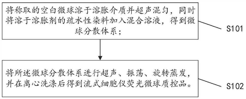
[0030] see figure 1 , the invention provides a method for preparing a flow cytometer fluorescent microsphere quality control product, comprising the following steps:

[0031] S101. Dissolving the weighed blank microspheres in the swelling medium and ultrasonically mixing, meanwhile, adding the hydrophobic dye dissolved in the swelling agent into the mixed solution to obtain a microsphere dispersion system.

[0032] Specifically, weigh 0.3 g to 1 g of blank microspheres with a coefficient of variation of forward scattered light less than 3%, and dissolve the blank microspheres in a swelling medium with a mass fraction of 1 to 5%, and use an ultrasonic intensity of 50 %~100% sonication for 1min~5min to obtain a mixed solution; weigh 2mg~10mg of hydrophobic dye and dissolve it in 20mL~50mL of swelling agent, add the mixed solution to obtain a microsphere dispersion system, The microspheres are polystyrene microspheres, the particle size should be suitable for flow cytometry, pre...

Embodiment 1

[0039]Weigh 0.3 g of blank microspheres with a particle size of 3.39 μm and a coefficient of variation of forward scattered light of 0.73%, dissolve them in 100 mL of Pluromic F127 aqueous solution (1% by mass fraction), and ultrasonically disperse them uniformly for 1 min (intensity is set to 50%) Weigh 2 mg of Nile Red dye and dissolve it in 20 mL of tetrahydrofuran, mix the two, ultrasonicate for 2 min (intensity is set to 100%), and vibrate on a constant temperature shaker for 30 min (room temperature is 21.3 °C, speed is 100 r / min), After the oscillation is completed, transfer to a rotary evaporator for rotary evaporation (temperature is set to 30°C, rotation speed is 60r / min, pressure is 0.05MPa, time is 10min), and the product after rotary evaporation is centrifuged (relative centrifugal force is 3000g, time is 2min) And washed repeatedly with ethanol until the supernatant was colorless. The supernatant was discarded, and the fluorescent microspheres were dispersed and ...

Embodiment 2

[0042] Weigh 0.5 g of blank microspheres with a particle size of 3.39 μm and a coefficient of variation of forward scattered light of 0.73%, dissolve them in 150 mL of Pluromic F127 aqueous solution (1% by mass fraction), and ultrasonically disperse them uniformly for 2 min (intensity is set to 50%) ; Dissolve 3.5mg of Nile Red dye in 35mL of tetrahydrofuran, mix the two, ultrasonicate for 3 minutes (intensity is set to 100%), shake on a constant temperature shaker for 40 minutes (room temperature is 22.8°C, speed is 120r / min) After the oscillation is completed, transfer to a rotary evaporator for rotary evaporation (the temperature is set to 37°C, the rotating speed is 60r / min, the pressure is 0.07MPa, and the time is 20min), and the product after the rotary evaporation is centrifuged (the relative centrifugal force is 3000g, and the time is 3min ) and washed repeatedly with ethanol until the supernatant was colorless. The supernatant was discarded, and the fluorescent micros...


PUM

PropertyMeasurementUnit
Particle sizeaaaaaaaaaa

Abstract

The invention discloses a flow cytometry fluorescent microsphere quality control product and a preparation method thereof. The flow cytometry fluorescent microsphere quality control product comprises 0.3-1 g of blank microspheres, 100-250 mL of a swelling medium, 2-10 mg of hydrophobic dye and 20-50 mL of a swelling agent. The method includes weighing 0.3-1 g of the blank microspheres with the forward scattering light variation coefficient smaller than 3%, dissolving the blank microspheres in the swelling medium with the mass fraction being 1-5%, carrying out ultrasonic treatment for 1-5 minutes by using an ultrasonic crusher with the ultrasonic intensity of 50-100%, weighing 2-10 mg of the hydrophobic dye, dissolving the hydrophobic dye in 20-50 mL of the swelling agent, and adding the mixed solution to obtain a microsphere dispersion system; and carrying out ultrasonic treatment, oscillation and rotary evaporation on the microsphere dispersion system, and carrying out centrifugal washing to obtain the flow cytometry fluorescent microsphere quality control product.

Description

technical field [0001] The invention relates to the technical field of fluorescent microspheres for flow cytometry, in particular to a fluorescent microsphere quality control product for flow cytometry and a preparation method thereof. Background technique [0002] Flow cytometer is a cell analyzer that measures the scattered light of cells and the intensity of labeled fluorescence. It can analyze tens of thousands of cells at high speed, and can simultaneously measure multiple cell characteristic parameters from one cell for qualitative and quantitative analysis. Fast, high precision, and good accuracy have become the most advanced cell quantitative analysis technology in the contemporary era and are widely used in clinical practice. At present, the quality control products of fluorescent microspheres for domestic flow cytometry all rely on imports, which are expensive. Although there are a small number of fluorescent microsphere standard substances suitable for flow cytom...

Claims


Application Information

Patent Timeline
IPC IPC(8): C09K11/02G01N15/14G01N1/30
CPCC09K11/025G01N15/14G01N1/30
Inventor 王丽钧张娟罗谢奥
Owner URIT MEDICAL ELECTRONICS CO LTD
Learn More