Establishment method of chronic ocular hypertension and long ocular axis animal model, model and application
A technology of animal models and establishment methods, applied in veterinary instruments, veterinary surgery, medical science, etc., can solve the problems of long model construction period, high economic cost, lack of simple and effective animal models, etc.
- Summary
- Abstract
- Description
- Claims
- Application Information
AI Technical Summary
Problems solved by technology
Method used
Image
Examples
Embodiment 1
[0053] This example is used to establish a chronic high eye pressure combined with long eye shaft animal model.
[0054] Preparation of model animals:
[0055] 40 SD healthy male rats were selected from 7-8 weeks, randomly divided into two groups, 20 each group, respectively as a model group and control group, respectively. In the temperature of 20-26 ° C, the relative humidity is 60%, and it is raised in an environment freely eating drinking water. 3 days before surgery, 8-10 o'clock per day, measured, was recorded as baseline intraocular pressure in the previous day, after which intraocular pressure detection was performed at this time period.
[0056] Establishment of animal models:
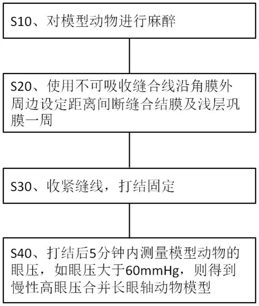
[0057] The establishment of animal models figure 1 As shown, mainly includes the following steps:
[0058] S10, anesthesia for model animals: 2% pentobarbital sodium is injected with 2% pentobarbital sodium in the abdominal cavity of the model group and the control group to perform a systemic anes...
Embodiment 2
[0069] The present embodiment uses the high eye pressure of the detection model animal and the change in the ball thereof.
[0070] 1 day, 1 week, 2 weeks, 4 weeks, February and June, and analyzed the changes of the intra-pressure meter in different time periods, and analyzed the eye pressure of the rat. Such as Figure 5 Indicated. by Figure 5 It can be seen that after the rat is sutured by a ring angle, the eye pressure suddenly increases when the suture is ligated. At this time, if Figure 4 The B is shown in the B figure, the corneal oxygen in the rats is white, corneal edema, and pupil. The rats under normal awake state were 10-13 mmHg, and the intraocular pressure of 5 minutes after the normal control group in this example was 12.2 ± 1.5 mmHg. The peak of the eye pressure measurement of the model group can reach 98 mmHg, and 5 minutes of intraoperative pressure dropped to 61.4 ± 10.4 mmHg. And gradually decline over time. The intraocular pressure obtained was 25.1 ± 3.6 mmHg 1...
Embodiment 3
[0074] This embodiment uses the long eye axis of the detection model animal and the change of the scleral tissue.
[0075]The model group rattles and control group rats were taken at 3 performed in the 4 weeks, and the complete eyeballs were taken out, and the complete eyeball was tagged. After the decada sclera (2mm after corneal), 2.5% glutaraldehyde fixation was fixed, prepared The electron microscope specimens use transmissive electron microstructures for ultrastructural observation of scleral collagen fibers. Such as Figure 12 As shown, the collagen fiber structure of the comparative control group in the model group ratio increased.
PUM
 Abstract
Description
Claims
Application Information
 - R&D
- Intellectual Property
- Life Sciences
- Materials
- Tech Scout
- Unparalleled Data Quality
- Higher Quality Content
- 60% Fewer Hallucinations
Browse by: Latest US Patents, China's latest patents, Technical Efficacy Thesaurus, Application Domain, Technology Topic, Popular Technical Reports.
© 2025 PatSnap. All rights reserved.Legal|Privacy policy|Modern Slavery Act Transparency Statement|Sitemap|About US| Contact US: help@patsnap.com


