
Kit for detecting cancer cells in ascites or abdominal cavity lavage fluid

A kit and lavage fluid technology, applied in the field of kits for detecting cancer cells in ascites or peritoneal lavage fluid, can solve the problems that conventional technologies cannot meet expectations, and achieve improved detection accuracy, high detection sensitivity, and sample saving amount of effect

Pending Publication Date: 2021-10-29
AFFILIATED HOSPITAL OF JIANGNAN UNIV +1
View PDF7 Cites 0 Cited by
  • Summary
  • Abstract
  • Description
  • Claims
  • Application Information

AI Technical Summary

Problems solved by technology

However, conventional techniques may not work as intended when only a small number of free cancer cells are present in the lavage fluid

Method used

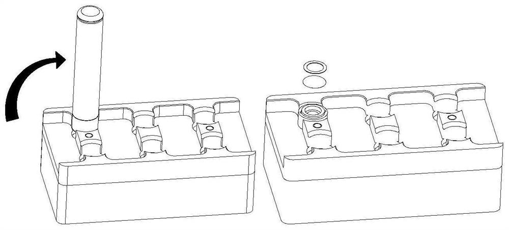
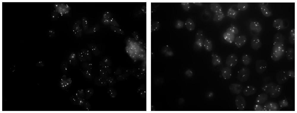
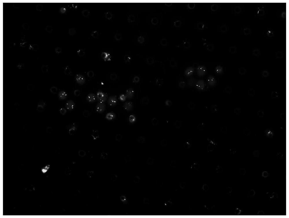

Image

  • Kit for detecting cancer cells in ascites or abdominal cavity lavage fluid
  • Kit for detecting cancer cells in ascites or abdominal cavity lavage fluid
  • Kit for detecting cancer cells in ascites or abdominal cavity lavage fluid

Examples


Embodiment 1

[0036] 1. The materials mainly involved in this embodiment are shown in Table 1.

[0037] Table 1 Main consumables and reagents

[0038]

[0039]

[0040] 2. Sample collection and rinsing

[0041] 1) Sample collection:

[0042] a) First collect 30mL ascites or peritoneal lavage fluid samples with a 50mL sterile tube.

[0043] b) The samples do not need to be refrigerated, room temperature is sufficient, and pretreatment (step 3) needs to be carried out within 2 hours after collection. If it cannot be handed over for testing within 2 hours, please store it vertically in a refrigerator at 4°C, and the storage time should not exceed 24 hours. Samples stored in a 4°C refrigerator should be rewarmed at room temperature for at least 30 minutes before processing.

[0044] 2) Rinse the filter

[0045] a) Open the upper and lower plugs of the filter and place the filter on the rinse stand.

[0046] b) Add about 2mL of 75% medical alcohol into the filter with a Pasteur tube ...


PUM


Abstract

The invention provides a kit for detecting cancer cells in ascites or abdominal cavity lavage fluid, and belongs to the technical field of peritoneal carcinoma disease detection. The kit comprises a nano microfiltration membrane filtering device used for collecting epithelial cells in abdominal cavity lavage fluid, an IF-FISH reagent, a fluorescence in situ hybridization reagent and an immunofluorescence staining reagent; to be detected markers comprise one or more of the following components: CEP8, CEP17 and EpCAM protein. The kit is specially used for ascites or abdominal cavity lavage fluid samples with a small amount of free cancer cells, and the detection sensitivity is high; meanwhile, two technologies of FISH and IF are adopted for dyeing and observation at the same time, so that the detection accuracy is improved, and the sample size is saved.

Description

technical field [0001] The invention belongs to the technical field of peritoneal cancer detection, in particular to a kit for detecting cancer cells in ascites or peritoneal lavage fluid. Background technique [0002] Gastric cancer, colorectal cancer, primary peritoneal cancer and other abdominal and pelvic malignant tumors progress locally to form tumors on the peritoneal surface, which is usually called peritoneal carcinomatosis (PC) (abbreviated as peritoneal cancer). Ascites or peritoneal lavage fluid cytology examination of free peritoneal cancer cells (cytology, CY) is the gold standard for the diagnosis of peritoneal micrometastases. Some literatures believe that free cancer cells in the peritoneal cavity are a prerequisite for the formation of peritoneal metastasis and an independent poor prognostic factor for gastric cancer (Lee SD, RyuKW, Eom BW, et al. Prognostic significance of peritoneal washing cytology patients with gastric cancer[J]. Br J Surg, 2012, 99(3)...

Claims


Application Information

Patent Timeline
IPC IPC(8): G01N33/574G01N21/64
CPCG01N33/57446G01N21/6428G01N2021/6432
Inventor 蔡东焱高其忠陆庭勋
Owner AFFILIATED HOSPITAL OF JIANGNAN UNIV
Learn More