Preparation method of biological 3D printing ink containing tissue protein compound

A tissue protein and 3D printing technology, which is applied in the field of bio-3D printing ink preparation, can solve the problems of complex action mechanism of bioactive factors, inability to reproduce the complexity of cells and their extramatrix in vivo, and achieve good clinical application prospects, high biological compatibility effect

Pending Publication Date: 2021-12-17
GENERAL HOSPITAL OF PLA
View PDF7 Cites 0 Cited by
  • Summary
  • Abstract
  • Description
  • Claims
  • Application Information

AI Technical Summary

Problems solved by technology

[0004] However, due to the wide variety of biologically active factors in the body, the distribution is different, and the mechanism of action is complex, the microenvironment created by simply adding one or several factors is far from the real in vivo environment, and cannot reproduce the cells and their extracellular matrix in vivo. complexity of

Method used


Image

  • Preparation method of biological 3D printing ink containing tissue protein compound
  • Preparation method of biological 3D printing ink containing tissue protein compound
  • Preparation method of biological 3D printing ink containing tissue protein compound

Examples


Embodiment 1

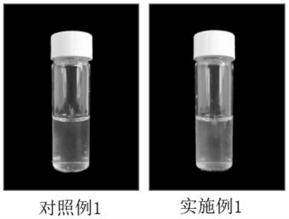
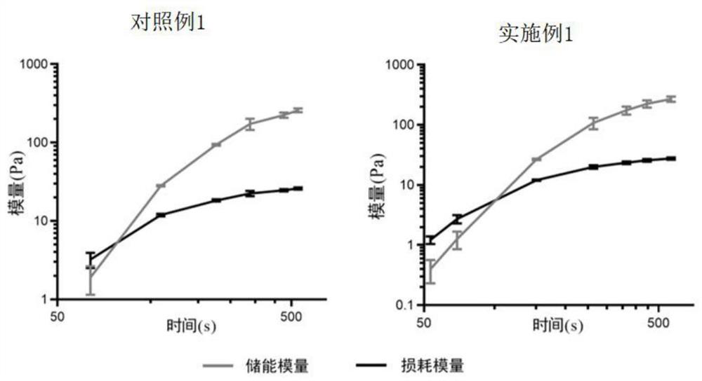
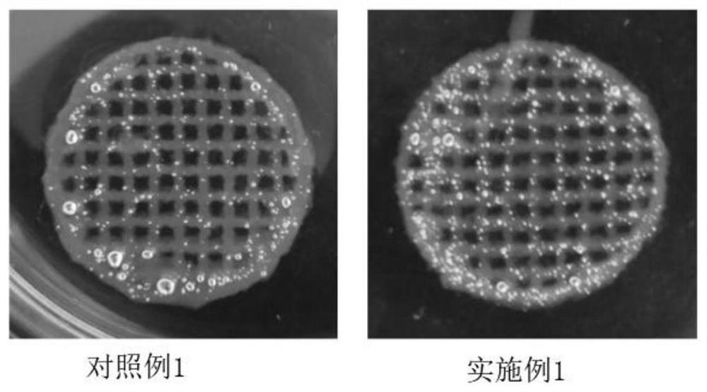
[0031] This embodiment provides a method for preparing a bio-3D printing ink containing a histone complex, which specifically includes the following steps:

[0032] Step S1 Tissue pretreatment: Take fresh human adipose tissue discarded from liposuction surgery, wash it with PBS several times until the washing solution is transparent and colorless, use ophthalmic scissors to remove fascia tissue, and cut the adipose tissue into about 0.1cm 3 For small pieces, take 10ml of the above-mentioned small pieces of adipose tissue, add 20ml of red blood cell lysate, lyse on ice for 15min, then pour the lysed pretreated tissue into a centrifuge tube, put it in a centrifuge at 2000rpm, 4 Under the condition of ℃, centrifuge for 5 minutes, take the upper fat tissue, and prepare the pretreated tissue;

[0033] Step S2 Extraction of tissue protein complexes: Place the pretreated tissue prepared in step S1 in a 20ml grinder for physical grinding until all the fat turns into a pale yellow chyl...


PUM

PropertyMeasurementUnit
wavelengthaaaaaaaaaa

Abstract

The invention provides a preparation method of biological 3D printing ink containing a tissue protein compound. The preparation method comprises the following steps: S1, tissue pretreatment: taking tissues, removing non-target components, cleaning with a PBS (Phosphate Buffer Solution), cutting the tissues into small blocks, cracking with a red blood cell lysis buffer, centrifuging, and taking supernatant so as to obtain the pretreated tissues; S2, extraction of the tissue protein compound: grinding the pretreated tissues by using a grinder, taking grinding liquid, carrying out secondary centrifugation, after layering the grinding liquid, taking an aqueous solution layer, carrying out tertiary centrifugation, taking supernate, and filtering by using a 0.22 mu m filter to obtain the tissue protein compound; and S3, preparation of the biological 3D printing ink containing the tissue protein compound: uniformly mixing the biological ink with the tissue protein compound to obtain the biological 3D printing ink. The preparation method has the beneficial effect that the biological 3D printing ink prepared by the invention has relatively high biocompatibility and biological reproducibility, meets the requirements of physiological or pathological mechanism exploration and tissue engineering repair, and has a relatively good clinical application prospect.

Description

technical field [0001] The invention relates to the field of medical model manufacturing and application, in particular to a preparation method of bio-3D printing ink containing tissue protein complex. Background technique [0002] 3D bioprinting technology is an emerging biomanufacturing technology, which simulates the microenvironment of tissues or organs in the body to the greatest extent by using cells and specific bioinks, and then manufactures 3D functional living tissues. Therefore, 3D bioprinting technology can be used to manufacture in vitro tissue / disease models, drug development and screening applications, understand disease development and progression, and fabricate tissues and organs for regenerative medicine, etc. [0003] Bio-ink is a key part of 3D bio-printing technology. In recent years, many natural or synthetic materials with good mechanical properties, biocompatibility and tissue regeneration have emerged, such as: gelatin, sodium alginate, methacryloyl ...

Claims


Application Information

Patent Timeline
Patent Type & Authority Applications(China)
IPC IPC(8): A61L27/36A61L27/22A61L27/20A61L27/02A61L27/50B33Y10/00B33Y70/10B33Y80/00
CPCA61L27/3687A61L27/3691A61L27/362A61L27/3604A61L27/3612A61L27/222A61L27/20A61L27/025A61L27/50B33Y10/00B33Y70/10B33Y80/00A61L2430/40A61L2300/412A61L2430/02C08L5/04
Inventor 张超李曌宋薇姚斌黄沙付小兵
Owner GENERAL HOSPITAL OF PLA
Why Eureka
Try Eureka