Microscopic slide preparation method for observing characteristics of lower surface of folium artemisiae argyi

A surface feature and microscopic preparation technology, which is applied in the preparation, sampling, and measuring devices of test samples to achieve the effects of high success rate, clear observation, and simple operation

Pending Publication Date: 2022-03-04
HENAN UNIV OF CHINESE MEDICINE
View PDF0 Cites 0 Cited by
  • Summary
  • Abstract
  • Description
  • Claims
  • Application Information

AI Technical Summary

Problems solved by technology

[0003] For above-mentioned situation, in order to overcome the deficiencies in the prior art, the object of the present invention is exactly to provide a kind of microscopic preparation method that is used to observe the lower surface feature of Folium Artemisiae Argyi, can effectively solve the problem that optical microscope or scanning electron microscope are easy to the lower surface of Folium Artemisiae Argyi. The distribution of stomata, glandular and non-glandular trichomes, and the pattern of the antiglandular wall of the epidermal cells.

Method used


Image

  • Microscopic slide preparation method for observing characteristics of lower surface of folium artemisiae argyi
  • Microscopic slide preparation method for observing characteristics of lower surface of folium artemisiae argyi
  • Microscopic slide preparation method for observing characteristics of lower surface of folium artemisiae argyi

Examples


Embodiment 1

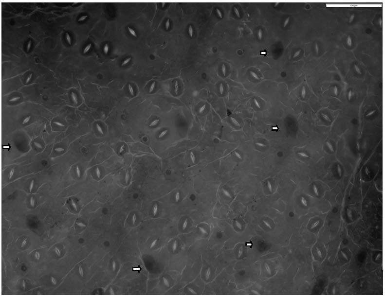
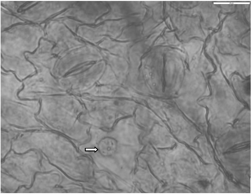
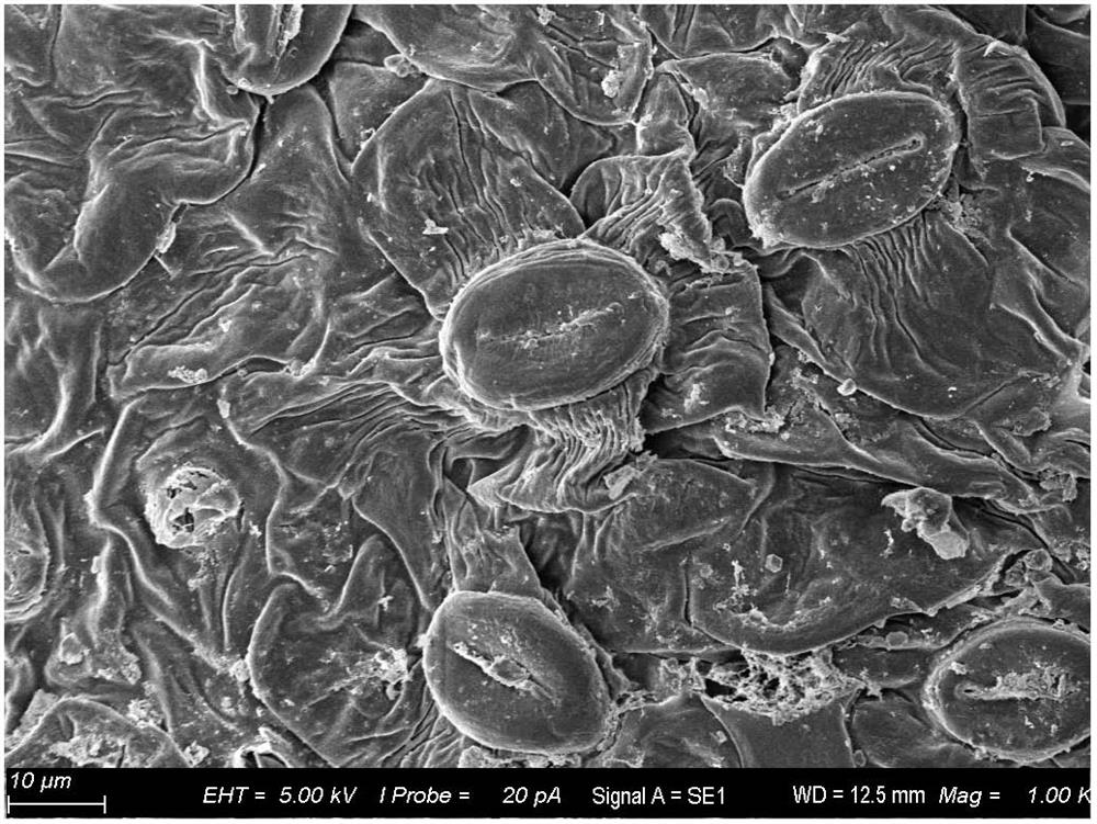
[0020] The present invention is a kind of microscopic preparation method for observing the lower surface feature of Folium Artemisiae Argyi, comprising the following steps:

[0021] (1) Blade glue: Take fresh moxa leaves, and evenly coat a thin layer of mixture of neutral glue and ceramic glue on the back of the leaf where the material is to be taken. The volume ratio of neutral glue and ceramic glue is 2:1. Stick a strip of paper on the mixed glue, press lightly to ensure the paste is firm;

[0022] (2) Peel off the non-glandular hairs: After the mixed glue dries, tear off the paper strips, the non-glandular hairs on the lower surface are peeled off from the lower surface and stick to the paper strips;

[0023] (3) Remove residual mixed glue: cut off a small piece of 7mm square leaves at the position where the non-glandular hairs are stripped, rinse in distilled water, and rinse repeatedly 4 times, remove the remaining mixed glue on the leaves, and get a clean small piece of ...

Embodiment 2

[0029] The present invention is a kind of microscopic preparation method for observing the lower surface feature of Folium Artemisiae Argyi, comprising the following steps:

[0030] (1) Blade glue: Take fresh moxa leaves, and evenly coat a thin layer of mixture of neutral glue and ceramic glue on the back of the leaf where the material is to be taken. The volume ratio of neutral glue and ceramic glue is 2:1. Stick a strip of paper on the mixed glue, press lightly to ensure the paste is firm;

[0031] (2) Peel off the non-glandular hairs: After the mixed glue dries, tear off the paper strips, the non-glandular hairs on the lower surface are peeled off from the lower surface and stick to the paper strips;

[0032] (3) Remove the residual mixed glue: cut off a small piece of 6mm square leaves at the position where the non-glandular hairs are stripped, rinse in distilled water, rinse repeatedly 3 times, remove the remaining mixed glue on the leaves, and get a clean small piece of ...

Embodiment 3

[0038] The present invention is a kind of microscopic preparation method for observing the lower surface feature of Folium Artemisiae Argyi, comprising the following steps:

[0039] (1) Blade glue: Take fresh moxa leaves, and evenly coat a thin layer of mixture of neutral glue and ceramic glue on the back of the leaf where the material is to be taken. The volume ratio of neutral glue and ceramic glue is 2:1. Stick a strip of paper on the mixed glue, press lightly to ensure the paste is firm;

[0040] (2) Peel off the non-glandular hairs: After the mixed glue dries, tear off the paper strips, the non-glandular hairs on the lower surface are peeled off from the lower surface and stick to the paper strips;

[0041] (3) Removal of residual mixed glue: at the position where the non-glandular hairs are peeled off, cut a small piece of leaf of 8mm square, rinse it in distilled water, and rinse it repeatedly for 5 times, remove the remaining mixed glue on the leaf, and get a clean sma...


PUM


Abstract

The invention relates to a microscopic slide preparation method for observing the characteristics of the lower surface of folium artemisiae argyi, and effectively solves the problem that an optical microscope or a scanning electron microscope is easy to observe the distribution of pores, glandular hairs and non-glandular hairs on the lower surface of the folium artemisiae argyi and the vertical peripheral wall pattern of epidermal cells. Small leaves are taken and rinsed with distilled water to remove residual gum, and part of the leaves are fixed with methanol, dehydrated with anhydrous alcohol, soaked with isoamyl acetate, dried at a critical point of carbon dioxide and sprayed with metal by an ion instrument for scanning electron microscope slide preparation observation; and soaking the other part of small leaves with a mixed solution of sodium hydroxide and sodium hypochlorite, dissociating mesophyll tissues, bleaching, rinsing with clear water, placing on a glass slide, dyeing with toluidine blue O, making a sodium iodide solution transparent, and sealing with water, thereby obtaining the optical microscope lens for observing stomata and epidermal cells by using a common optical microscope or fluorescence microscope. The method is simple to operate, low in cost, small in workload, high in success rate, clear in observation and good in effect.

Description

technical field [0001] The invention relates to the field of microscopic identification of crude drugs, in particular to a microscopic preparation method for observing the characteristics of the lower surface of mugwort leaves, which can observe the characteristics of the leaf epidermis by treating the lower surface of the leaves of mugwort leaves. Background technique [0002] Artemisia argyi is a commonly used traditional Chinese medicine, and it is produced in most parts of the country. Artemisia argyi leaves are dried and crushed to obtain "Moxa velvet", which is used to make moxa sticks for moxibustion. Some characteristics of the surface of Artemisia argyi, such as the type and density of the indumentum, stomatal type, stomatal index, and anticlinal wall pattern of epidermal cells, are not only of important reference value for the identification of Artemisia argyi, but also for the study of its plant ecology, plant physiology and variety selection. also plays an impor...

Claims


Application Information

Patent Timeline
Patent Type & Authority Applications(China)
IPC IPC(8): G01N1/28
CPCG01N1/2813
Inventor 郭涛冯书营雷青会许二平回成程刘孟奇张振强
Owner HENAN UNIV OF CHINESE MEDICINE
Why Eureka
Try Eureka