
Method and device for performing tumor diagnosis by using ultrasonic image fusion data

A technology of tumor diagnosis and ultrasound image, applied in the field of breast detection, can solve the problems of high missed diagnosis rate, time-consuming, unsuitable for early screening methods, etc., and achieve the effect of improving efficiency

Pending Publication Date: 2022-03-15
浙江衡玖医疗器械有限责任公司
View PDF0 Cites 0 Cited by
  • Summary
  • Abstract
  • Description
  • Claims
  • Application Information

AI Technical Summary

Problems solved by technology

[0004] Among them, mammography is generally used as the gold standard in breast imaging examination, but the process of mammography examination needs to compress the breast, especially for the evaluation of suspicious breast calcification in dense breasts, the specificity is not high, and the missed diagnosis rate is as high as 76%
Compared with Western women, Eastern women's breasts are relatively dense, and the age of onset of breast cancer is lower. Therefore, mammography is not suitable as an early screening method for Asian women's breast examination; traditional ultrasound examination can only provide images of a certain section of breast tissue , in clinical practice, doctors need to spell out the three-dimensional structure of breast tissue in their minds based on real-time two-dimensional images, which is highly subjective, lacks repeatability and traceability, and it is easy to produce inaccurate estimates for the location, shape and size of tumors ; Although nuclear magnetic resonance has the advantage of sensitivity, it is time-consuming and expensive, and is not suitable as an early screening method; and improved ultrasonic inspection equipment usually includes GE's ABUS and Siemens' ABVS. Although these improved ultrasonic inspection equipment can obtain three-dimensional images , but only collect the reflected wave signal to form a gray-scale image, which shows a relatively simple presentation of the breast tissue, and cannot clearly show the obvious difference between the lesion location and the normal breast tissue

Method used


Image

  • Method and device for performing tumor diagnosis by using ultrasonic image fusion data
  • Method and device for performing tumor diagnosis by using ultrasonic image fusion data
  • Method and device for performing tumor diagnosis by using ultrasonic image fusion data

Examples


Embodiment Construction

[0028] The technical solutions in the embodiments of the present invention will be clearly and completely described below in conjunction with the accompanying drawings in the embodiments of the present invention. Obviously, the described embodiments are only some embodiments of the present invention, not all embodiments. All other embodiments obtained by persons of ordinary skill in the art based on the embodiments of the present invention belong to the protection scope of the present invention.

[0029] Those skilled in the art should understand that, in the disclosure of the present invention, the terms "vertical", "transverse", "upper", "lower", "front", "rear", "left", "right", " The orientation or positional relationship indicated by "vertical", "horizontal", "top", "bottom", "inner", "outer", etc. is based on the orientation or positional relationship shown in the drawings, which are only for the convenience of describing the present invention and The above terms should ...


PUM


Abstract

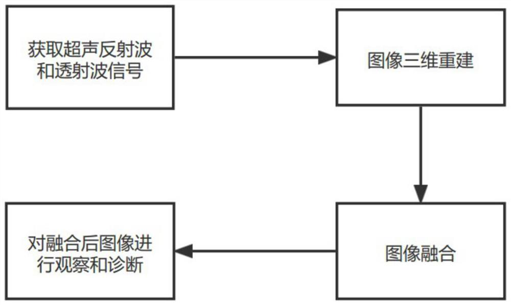
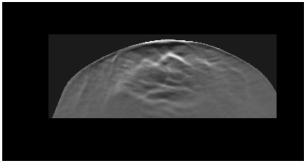
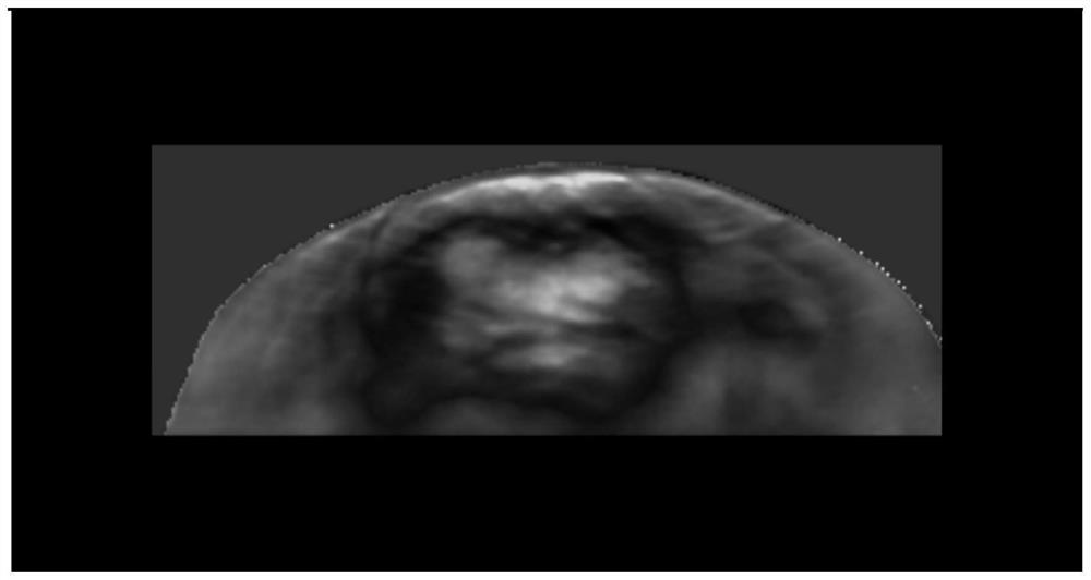
The invention provides a method for carrying out tumor diagnosis by utilizing ultrasonic image fusion data, which comprises the following steps of: transmitting ultrasonic waves to mammary gland tissues to obtain reflected wave signals and transmitted wave signals aiming at different directions of the mammary gland tissues, and covering all-directional angles of the mammary gland tissues in the different directions; performing image reconstruction on the obtained reflected wave signal and transmitted wave signal to obtain a sound velocity image, a reflected image and an attenuated image; performing image fusion on the sound velocity image, the reflection image and the attenuation image to obtain a fused image; the three-dimensional ultrasonic image data corresponding to the mammary gland tissue can be generated only through one-time scanning, some key information can be displayed in an overlapped mode after the three images are fused, imaging performance of different signals on the mammary gland tissue can be displayed on the same image, and doctors can find specific performance of diseases easily.

Description

technical field [0001] The invention relates to the field of mammary gland detection, in particular to a method and device for tumor diagnosis using ultrasonic image fusion data. Background technique [0002] Breast cancer is the most common cancer in the world. In 2020, according to the World Health Organization’s International Agency for Research on Cancer, breast cancer has 2.26 million new cancer cases, surpassing lung cancer and becoming the world’s most common cancer. In China, breast cancer is even more It is the number one killer of women's health, and compared with Europe and the United States, due to the lack of early screening methods, the spread of cancer cells is often found when it is discovered. [0003] Existing examination methods for breast cancer generally include mammography, traditional ultrasonography, nuclear magnetic resonance, and modified ultrasonography. [0004] Among them, mammography is generally used as the gold standard in breast imaging exam...

Claims


Application Information

Patent Timeline
IPC IPC(8): A61B8/00A61B8/08
CPCA61B8/5215A61B8/0825A61B8/5207A61B8/44A61B8/463
Inventor 胡雪燕陈慧君朱坚栋黄跃龙
Owner 浙江衡玖医疗器械有限责任公司
Learn More