Construction method and application of urinary tract contrast animal model based on cystostomy

A technology of urography and animal models, which is applied in the field of urography animal models to achieve the effect of promoting research

Pending Publication Date: 2022-07-15
谭硕 +1
View PDF0 Cites 0 Cited by
  • Summary
  • Abstract
  • Description
  • Claims
  • Application Information

AI Technical Summary

Problems solved by technology

[0006] Establishing an ideal experimental animal model of the urinary system is very important for the study of the development and molecular mechanism of human urinary system-related diseases, but there is no animal model of urography through cystostomy for clinical research

Method used


Image

  • Construction method and application of urinary tract contrast animal model based on cystostomy
  • Construction method and application of urinary tract contrast animal model based on cystostomy
  • Construction method and application of urinary tract contrast animal model based on cystostomy

Examples


Embodiment 1

[0036] (1), weigh and anesthetize the rat according to a certain drug dose; after the rat enters an anesthetized state, after shaving around the lower abdomen of the rat to prepare the skin, use complex iodine solution to disinfect the lower abdomen and external urethra;

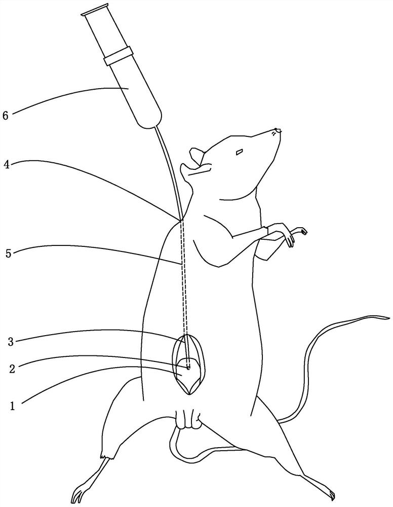
[0037] (2), under aseptic conditions, cut the skin and the rectus abdominis layer by layer along the midline of the abdomen of the rat, expose the bladder, puncture the ventral midline of the bladder with a surgical blade to form a cystostomy opening, and drain the urine in the bladder liquid, and insert a cystostomy tube made of a venous blood collection needle hose into the cystostomy opening;

[0038] (3) A 2mm skin puncture hole was made 2cm below the line connecting the upper edges of the two ears at the back of the rat's neck with a surgical blade in advance, and a hemostatic forceps were used to bluntly separate the upper end of the bladder along the subcutaneous plane to the back of the neck skin punc...

Embodiment 2

[0042] (1), weigh and anesthetize the rat according to a certain drug dose; after the rat enters an anesthetized state, after shaving around the lower abdomen of the rat to prepare the skin, use complex iodine solution to disinfect the lower abdomen and external urethra;

[0043] (2), under aseptic conditions, cut the skin and the rectus abdominis layer by layer along the midline of the abdomen of the rat, expose the bladder, puncture the ventral midline of the bladder with a surgical blade to form a cystostomy opening, and drain the urine in the bladder liquid, and insert a cystostomy tube made of a venous blood collection needle hose into the cystostomy opening;

[0044] (3) In advance, a 3mm skin puncture hole was made 3cm below the line connecting the upper edges of the two ears at the back of the rat's neck with a surgical blade, and a hemostatic forceps were used to bluntly separate the upper end of the bladder along the subcutaneous plane to the back of the neck skin punct...

Embodiment 3

[0048] (1), weigh and anesthetize the rat according to a certain drug dose; after the rat enters an anesthetized state, after shaving around the lower abdomen of the rat to prepare the skin, use complex iodine solution to disinfect the lower abdomen and external urethra;

[0049] (2), under aseptic conditions, cut the skin and the rectus abdominis layer by layer along the midline of the abdomen of the rat, expose the bladder, puncture the ventral midline of the bladder with a surgical blade to form a cystostomy opening, and drain the urine in the bladder liquid, and insert a cystostomy tube made of a venous blood collection needle hose into the cystostomy opening;

[0050] (3) A 4mm skin puncture hole was made 1cm below the line connecting the upper edges of the two ears at the back of the rat's neck with a surgical blade in advance, and a hemostatic forceps were used to bluntly separate the upper end of the bladder along the subcutaneous plane to the back of the neck skin punc...


PUM


Abstract

The invention belongs to the technical field of medical animal models, and relates to a bladder fistulization model, which is characterized in that under a sterile condition, a surgical blade is used for puncturing a bladder to form a bladder fistulization opening, urine in the bladder is drained, a bladder fistulization tube is placed in the bladder fistulization opening, the bladder fistulization opening is sutured by an absorbable surgical suture, and a skin puncture hole is formed in the rear part of the neck of a rat. A subcutaneous tunnel is formed from the upper end plane of the bladder to the subcutaneous tissue of the skin puncture hole, and the tail end of the bladder fistulization tube penetrates through the rectus abdominis to the subcutaneous part and then penetrates out of the skin puncture hole along the subcutaneous tunnel. When bladder and urethra angiography is carried out smoothly, an injector containing a contrast agent is connected with the tail end of the bladder fistulization tube, the contrast agent is injected to display the whole process of the bladder and the urethra of the rat, and the rat animal model greatly promotes research on the urinary system of the rat and can be applied to other animals for reference at the same time. The method has great significance in research on clinical related diseases of the human urinary system.

Description

technical field [0001] The invention belongs to the technical field of medical animal models, and in particular relates to a construction method and application of a urography animal model based on cystostomy. Background technique [0002] The bladder is an important urinary storage organ in mammals. After the urine is produced by the kidneys, it is transported into the bladder by the peristalsis of the ureter, and temporarily stored. When the bladder stores urine to a certain capacity, due to the increase of pressure, the stretch receptors in the bladder wall are excited by the stimulation, and the impulse is transmitted through the bladder. The nerve reaches the lower urination center in the lumbar spinal cord. At the same time, the spinal cord uploads the information of bladder filling to the advanced voiding center of the cerebral cortex, and produces the urge to urinate. After preparing to urinate, the brain sends nerve impulses along the descending conduction tract t...

Claims


Application Information

Patent Timeline
Patent Type & Authority Applications(China)
IPC IPC(8): A61D1/00A61D7/00
CPCA61D1/00A61D7/00
Inventor 谭硕吴自强唐正严王钊朱泉丁科李恺璇刘彦伶
Owner 谭硕
Why Eureka
Try Eureka