Protein chip for detecting blood and cerebro spinal fluid pathogen antibody, and its preparing method and use

A pathogen antibody and protein chip technology, which is applied in biochemical equipment and methods, measuring devices, and microbial determination/inspection. Large and other problems, to achieve the effect of retaining a high degree of specificity, improving detection speed and efficiency, and using less sample

Inactive Publication Date: 2007-02-14
SHANDONG MEDICAL BIO TECH RES CENT
View PDF10 Cites 0 Cited by
  • Summary
  • Abstract
  • Description
  • Claims
  • Application Information

AI Technical Summary

Problems solved by technology

[0006] For herpes simplex virus type I (HSV-I), herpes simplex virus type II (HSV-II), varicella-zoster virus (VZV), cytomegalovirus (CMV), Epstein-Barr virus (EBV), rubella virus (RV), Japanese encephalitis virus (JEV), mumps virus (MV), Mycobacterium tuberculosis, and Leptospira to detect ten kinds of pathogens, it takes 20 kits to detect all IgG and IgM, Each reaction can only detect one indicator, which is slow, inefficient, expensive, and requires a large amount of samples, which is actually impossible; moreover, the antigens in the currently used detection kits are all crude antigens extracted from pathogen cultures. The virus content is low, the composition is complex, the background is high, the cross-reaction between various pathogens is serious, and the specificity, sensitivity, and stability vary greatly; in addition, the substrates used in the experiment are also highly toxic chemicals. There is also a potential threat to the health of personnel

Method used


Image

  • Protein chip for detecting blood and cerebro spinal fluid pathogen antibody, and its preparing method and use
  • Protein chip for detecting blood and cerebro spinal fluid pathogen antibody, and its preparing method and use
  • Protein chip for detecting blood and cerebro spinal fluid pathogen antibody, and its preparing method and use

Examples


Embodiment 1

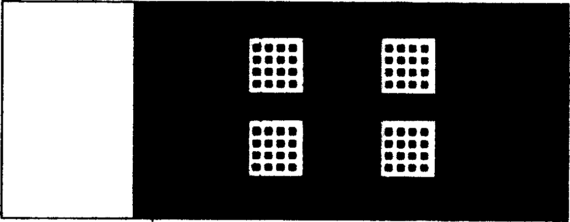
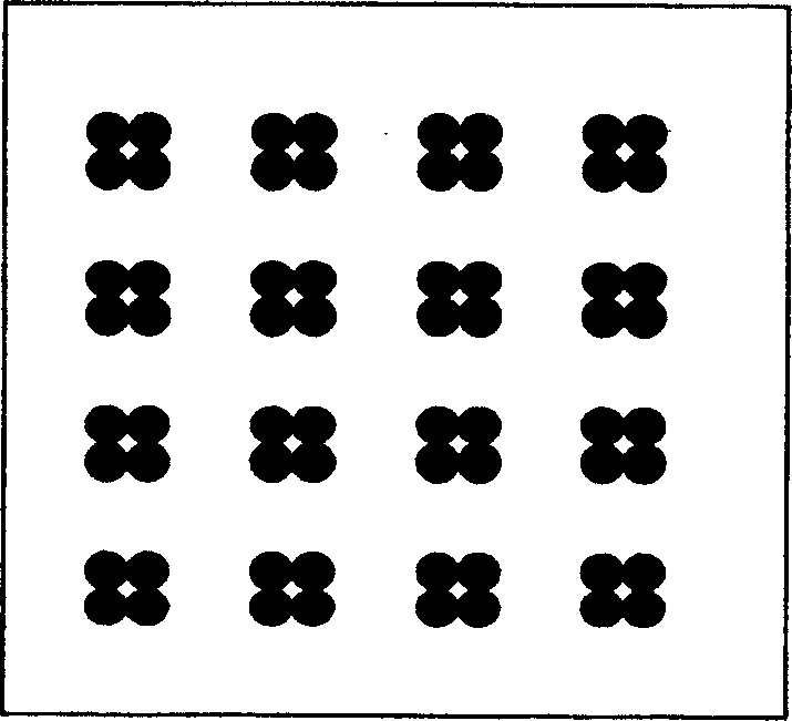
[0046] Example 1: Modification of slides and immobilization of antigens, such as figure 1 To detect the appearance of the antibody protein chip of serum cerebrospinal fluid pathogens, such as figure 2 Spot array diagram.

[0047] Place the slide on the slide rack, put it into a glass jar containing 350ml cleaning solution (NaOH 100g, ethanol 600ml, water 400ml), place it on a shaker at 60 rpm, shake it for 2 hours; discard the cleaning solution, Wash 4 times with water for 3 minutes each time; soak the slides in a glass jar filled with 350ml polylysine PBS solution (polylysine 35ml, PBS 35ml, water 280ml), and set a shaker at 60 rpm , Shake for 1 hour; immerse the slides in water and wash 5 times up and down; put in a centrifuge, 800 rpm / 5 minutes after separating the core, put it in a clean plastic box, place it vertically for 2 weeks or place the sample after baking slices in the oven use.

[0048] Do 2 serum samples testing, choose 2×2 microarray chip, (such as figure 1 ) ...

Embodiment 2

[0050] Example 2: Antigen-antibody reaction and detection

[0051] Add blocking solution (1% BSA, 0.2g / L KCl, 1.44g / L Na2HPO4, 0.24g / L KH2PO4, 8g / L NaCl, 0.1% Tween-20,) to block on the chip with good antigen, 37℃1 Hours, block non-specific sites on the substrate surface; wash with PBST (0.2g / L KCl, 1.44g / L Na2HPO4, 0.24g / L KH2PO4, 8g / L NaCl, 0.1% Tween-20) 3 times, 10 times each time After seeding seconds, rinse with PBS (0.2g / L KCl, 1.44g / L Na2HPO4, 0.24g / L KH2PO4, 8g / L NaCl), centrifuge at 800 rpm / centrifuge for 3 minutes to remove excess blocking solution; Dilute 10 times with PBS and add 3μL to the array. The first sample is added to the upper and lower arrays in the first column; the second sample is added to the upper and lower arrays in the second column and placed in the hybridization box at 37℃ 30 minutes to make the antigen and antibody fully react; wash 3 times with PBST, after 10 seconds each time, rinse with PBS, centrifuge at 800 rpm / centrifuge for 3 minutes to remo...


PUM


Abstract

The invention discloses a protein chip for detecting pathogen antibodies of blood and cerebrospinal fluid and its preparing method, and the protein chip includes substrate and peculiar-culture or shape protein or polypeptide antigen of array distributed pathogen and contrasted dot coating, where the substrate is a glass substrate; and the antigen and contrasted dot coating refers to 13 antigens with ten indexes including HSV-I, HSV-II, VZV, CMV, EBV, RV, JEV, MV, MT and LP, uniformly distributed on the glass substrate in a dot matrix form, and positive contrast, negative contrast and blank contrast. The protein chip of the invention can obtain multiple-index reacting result only by one reaction, judges infection of different pathogens and has the characters of quickness, high efficiency, accuracy and parallel diagnosis.

Description

Technical field [0001] The invention relates to a biochip and a preparation method and application thereof, in particular to a low-density protein chip and a preparation method and application thereof, in particular to a blood cerebrospinal fluid pathogen antibody detection chip and a preparation method and application thereof. Background technique [0002] In recent years, the incidence of central nervous system (CNS) infectious diseases has increased. The pathogens include various bacteria, fungi, viruses, spirochetes, etc., and pathogenic diagnosis is the gold standard for diagnosis. Once the etiological diagnosis is confirmed, there is no need to mention the differential diagnosis. At present, pathogen diagnosis still relies on morphological examination, pathogen isolation and culture, and enzyme-linked immunosorbent assay (Enzyme-linked immunosorbent assay, ELISA) to detect antibodies; and for tuberculosis, spirochetes and various viral infections, including herpes simplex v...

Claims


Application Information

Patent Timeline
Patent Type & Authority Patents(China)
IPC IPC(8): G01N33/569G01N33/533C12Q1/70G01N33/552
Inventor 韩金祥刘毅
Owner SHANDONG MEDICAL BIO TECH RES CENT
Why Eureka
Try Eureka