High affinity immune globulin binding molecule and method for preparation
A technology that combines molecules and affinity, applied in the fields of molecular biology and immunodiagnostics
- Summary
- Abstract
- Description
- Claims
- Application Information
AI Technical Summary
Problems solved by technology
Method used
Image
Examples
Embodiment 1
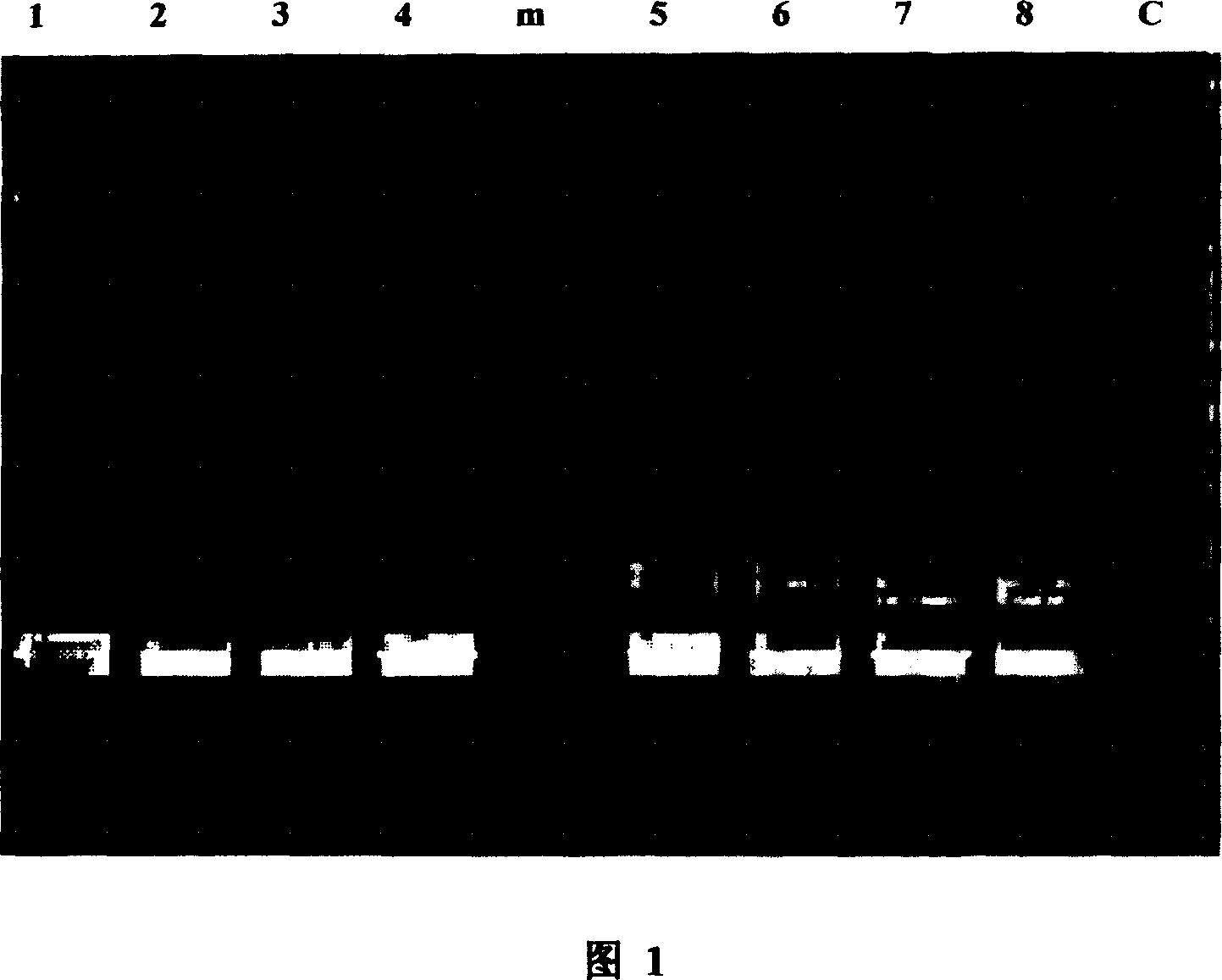
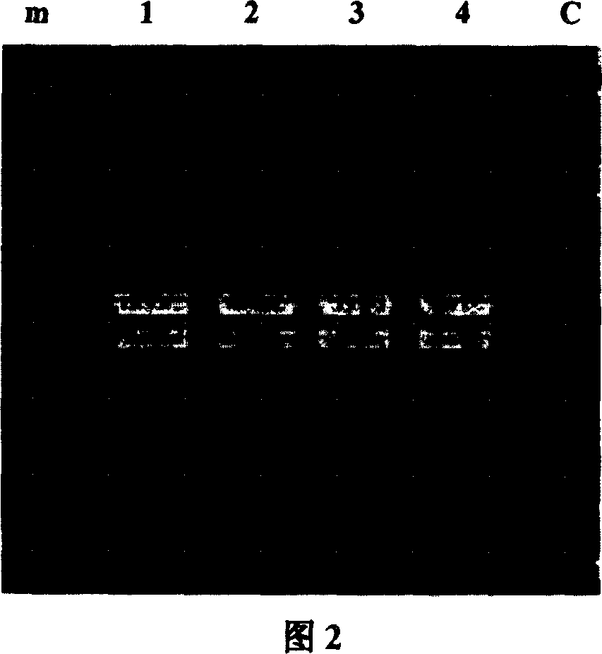
[0082] Example 1 Construction of Recombinant Ig Binding Molecule Library
[0083] 1. Synthesize primers
[0084] 1.1 For protein A, protein L, protein G single domain gene amplification
[0085] (1) The upstream primers for amplifying the sequence of Protein A-A, Protein A-B / C, Protein A-D, and Protein A-E from Mu-protein A / pGEM-T easy plasmid are:
[0086] PA-uA: 5’-cacgagctcGCTGACAACAATTTCAAC-3’
[0087] PA-uB: 5’-cgcgagctcGCGGATAACAAATTCAAC-3’
[0088] PA-uC: 5’-cgcgagctc GCTGACAACAAATTCAAC-3’
[0089] PA-uD: 5’-tccgagctcGCTGATGCGCAACAAAAT-3’
[0090] PA-uE: 5’-cgcgagctcGCTCAACAAAATGCTTTT-3’
[0091] The downstream primers are:
[0092] PA-dA: 5’-tctgagctcTTTCGGTGCTTGAGATTC-3’
[0093]PA-dB: 5’-tctgagctcTTTTGGTGCTTGTGCATC-3’
[0094] PA-dC: 5’-acggagctcTTTTGGTGCTTGAGCATC-3’
[0095] PA-dD: 5’-tcggagctcAAAGCCACGAACTCTAAG-3’
[0096] PA-dE: 5’-agagagctcTTTTGGAGCTTGAGAGTC-3’
[0097] (2) Amplify the B1-B5 sequence of Protein L from the Protein L / pGEM-T easy plasmid
[0098] The up...
Embodiment 2
[0131] Example 2. Phage display of recombinant Ig binding molecule library
[0132] 1. Preparation and linearization of phagemid vector pCANTAB5S DNA:
[0133] The pCANTAB5S phagemid DNA was extracted with the plasmid recovery kit of Shenneng Group, and proceeded according to the instructions. Quantification by agarose electrophoresis. After Sac I digestion, the alkaline phosphatase (CIAP) is dephosphorylated, and the linearized vector is recovered from the solution by the kit, and quantified by agarose electrophoresis.
[0134] 2. The Ig binding molecule library is connected to the phagemid vector:
[0135] The 12 kinds of recovered protein A fragments with single binding domains A, B, C, D, E, B1, B2, B3, B4, B single binding domain fragments of protein L, G1, G2 single binding domains of Protein G 80μl (5μg) of the mixture of binding domain digested fragments plus 5μl (25U) of T4 DNA ligase, 1.5μl of 10×Buffer, and a total reaction volume of 100μl. Divide 100μl into 20μl / branch...
Embodiment 3
[0142] Example 3. In vitro directed molecular evolution of Ig to screen high-affinity Ig binding molecules
[0143] 1. Ig affinity screening of recombinant phage library:
[0144] Coat 200μl of pH 9.6 carbonate buffer with human Ig (final concentration of 10μg / ml) on the enzyme-labeled strip plate at 37°C for 2 hours, and block the strip with 150μl of blocking solution (PBS+10% skimmed milk) Plate for 1 hour and store at 4°C. After washing 5 times with washing solution (PBS+0.05% Tween20), it was used for affinity screening of phage library. Add 100μl of blocking solution and 100μl of PALGn recombinant phage to each well of the slatted plate, react at 37°C for 3 hours, wash with washing solution (0.25% Tris+0.05% Tween20) 30 times, add 100μl of E. coli TG1 in logarithmic growth phase, react at 37°C for 1 After collecting, take 10μl, 1μl, 0.1μl coated with LB (containing ampicillin 100ng / ml) plates and count them overnight at 37°C to evaluate the binding of the phage library to Ig ...
PUM
 Abstract
Description
Claims
Application Information
 - R&D
- Intellectual Property
- Life Sciences
- Materials
- Tech Scout
- Unparalleled Data Quality
- Higher Quality Content
- 60% Fewer Hallucinations
Browse by: Latest US Patents, China's latest patents, Technical Efficacy Thesaurus, Application Domain, Technology Topic, Popular Technical Reports.
© 2025 PatSnap. All rights reserved.Legal|Privacy policy|Modern Slavery Act Transparency Statement|Sitemap|About US| Contact US: help@patsnap.com


