HEV antigenic peptide and its method

A technology of antigens and antibodies, applied in chemical instruments and methods, botany equipment and methods, biochemical equipment and methods, etc., can solve problems such as difficulties in breeding viruses

Inactive Publication Date: 2003-01-15
BEIJING WANTAI BIOLOGICAL PHARMACY ENTERPRISE
View PDF0 Cites 5 Cited by
  • Summary
  • Abstract
  • Description
  • Claims
  • Application Information

AI Technical Summary

Problems solved by technology

But so far it has not been possible to use live attenuated or killed virus particles to prepare a vaccine against HEV infection because of the great difficulty of propagating the virus in cultured cells

Method used


Image

  • HEV antigenic peptide and its method
  • HEV antigenic peptide and its method
  • HEV antigenic peptide and its method

Examples


Embodiment 1

[0171] Serum—96 parts of serum described in the present invention are obtained from non-A, non-B, and non-C acute hepatitis patients who are onset or have been ill with the permission of Hong Kong Queen Margaret Hospital. Samples were collected from patients who were hospitalized or at various times after discharge. The extraction of the cloning HEV RNA of embodiment 1 HEV capsid gene

[0172] RNA was extracted from the liver juice of rhesus monkeys experimentally infected with HEV (Zhuang et al., 1992) using the QIAamp Virus RNA kit (QIAGEN GbmH, Hilden, Germany) according to the manufacturer's instructions. The purified RNA was mixed with two volumes of isopropanol (BDH Laboratory Supplies, England), and 1 / 10 volume of 3M sodium acetate (pH6.4) (Sigma, USA), and placed at -20°C for 1 hour. Afterwards, the mixture was centrifuged at 15,000 rpm for 15 minutes. The pellet was washed once with 70% ethanol (BDH Laboratory Supplies, UK), and then resuspended in reverse transcrip...

Embodiment 2

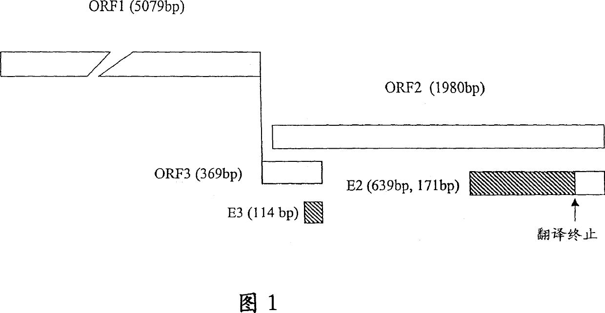
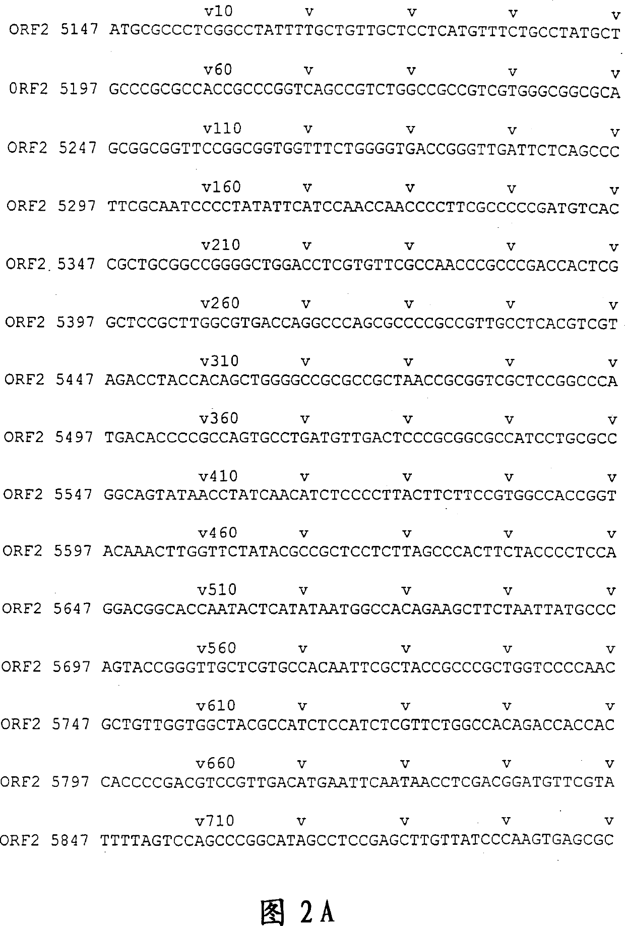
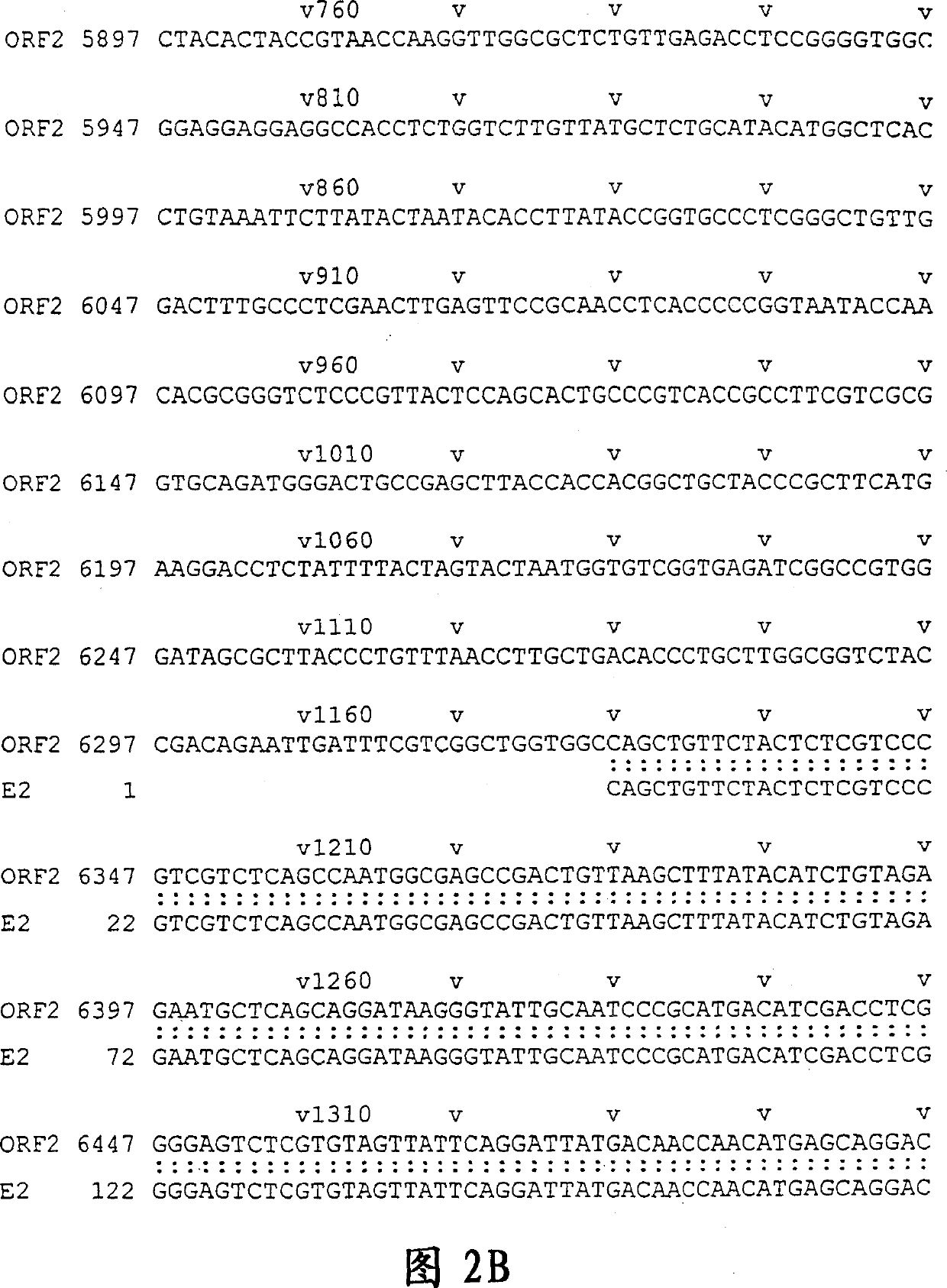
[0179] DNA sequencing was performed with ABI PRISM Dye Terminator Cycle Sequencing Ready Reaction Kit [PERKIN ELMER]. The results showed that the 811bp E2 sequence was located at positions 6326-7136, with a single base deletion at position 6957, presumably caused by PCR amplification errors. The resulting frameshift mutation caused premature termination of translation at a new stop codon at position 6968, resulting in a smaller-than-predicted 213 amino acid polypeptide with a molecular weight of 23 kD, rather than the originally expected 267 amino acids. The location of E2 and the associated fragments are shown in Figure 1. Example 2 Production of HEV Polypeptide and Expression of Purified GST Fusion Protein

[0180] The recombinant plasmid was transformed into BL21 E. coli. Pick a single clone in 2×YTA medium (peptone 16g / L; yeast extract 10g / L; NaCl 5g / L; penicillin 100ug / L). Inoculate 4ml of the overnight culture into 400ml of 2×YTA medium and culture at 28°C until OD600...

Embodiment 3

[0182] Thrombin [Pharmacia Biotech, USA] (5 ul of 1 U / ul solution) and 95 ul of PBS were added to 100 ul of GE2 bound to Glutathione Gel-4B and incubated at 22°C for 16 hours. The supernatant was collected by centrifugation, and the supernatants from the second substrate wash were pooled together. This protein cut out by thrombin is named pE2. Example 3 Identification of HEV polypeptide SDS PAGE (SDS polyacrylamide gel electrophoresis) analysis

[0183] 10% SDS polyacrylamide gels were prepared according to standard methods (Sambrook et al., 1989). 4ug polypeptide samples were loaded onto the gel, and electrophoresis was performed at 100 volts for 3 hours with a Minigel TwinG42 [Biometra, Germany] electrophoresis tank. The gel was stained with a Coomassie brilliant blue R250 mixture (45 ml methanol, 45 ml water and 10 ml glacial acetic acid). Western blot

[0184] After the SDS-PAGE, use the Mini-PROTEIN II Cell electrotransfer instrument [BIO-RAD] for one hour at 100 volt...


PUM


Abstract

A highly immunoreactive viral peptide, pE2, is disclosed which is derived from the carboxy-terminal end region of ORF2 region of the hepatitis E virus (HEV) genome. A unique feature of the novel pE2 peptide is that it possesses conformational antigenic determinants which are only exposed when monomers of the peptide associate with one another through non-covalent interactions to naturally form homodimers. The novel pE2 peptide is proven to be highly reactive with sera from patients having current or past infection with HEV which suggests that the homodimer may mimic certain structural features of the HEV capsid protein. Furthermore, the antigenic activity of the pE2 peptide is strictly conformational in nature and therefore, exhibits immunochemical reactivity only when the peptide exists in a dimeric form. Consequently, the antigenic activity is lost upon dissociation of the dimers, but the activity is restored when the monomers reassociate to form dimers. Moreover, diagnostic methods useful in detecting and diagnosing HEV infection, and the use of a vaccine composition effective in preventing hepatitis E virus infection in which the novel pE2 peptide is utilized are also disclosed.

Description

[0001] The present invention relates to a highly immunoreactive viral peptide pE2 cloned from the genome of a Chinese strain of hepatitis E virus (HEV), and its use in the development of reliable diagnostic methods for the detection and diagnosis of HEV, and in the prevention of human Application of HEV vaccine composition. Background of the invention [0002] Hepatitis E virus (HEV), first discovered in 1983, causes enterically transmitted hepatitis (Balayan et al., 1983). In 1991, the full-length genome of the virus was cloned and sequenced for the first time, and it was found to be a single-stranded positive sense non-enveloped RNA (Tam et al., 1991). Although HEV is morphologically similar to members of the Caliciviridae family (Bradley et al., 1988; Huang et al., 1992; Panda et al., 1989), it has a unique genomic organization (Berke et al., 1997). Based on sequence analysis, it is speculated that the 7.2 kb viral genome contains three open reading fram...

Claims


Application Information

Patent Timeline
Patent Type & Authority Applications(China)
IPC IPC(8): G01N33/564A61K38/00A61K39/00A61P31/20C07K14/08C07K16/10C12N1/15C12N1/19C12N1/21C12N5/10C12N9/10C12N15/09C12N15/51C12P21/02C12Q1/68G01N33/576
CPCY10S435/975C07K2319/00C07K16/10C07K2319/003G01N33/5767A61K39/00C12N2770/28122C07K14/005A61P31/14A61P31/20C07K14/08
Inventor 吴文翰严弘杰张纪忠
Owner BEIJING WANTAI BIOLOGICAL PHARMACY ENTERPRISE
Why Eureka
Try Eureka