Application of microdrop control in virus detection and detection method and chip

A virus detection and microdroplet technology, applied in the field of biochemical detection, can solve the problems of low detection accuracy and reliability, long detection period, small amount of detection, etc., and achieve the effects of low power consumption, low cost, and low sample consumption.

Inactive Publication Date: 2007-02-14
TSINGHUA UNIV
View PDF0 Cites 22 Cited by
  • Summary
  • Abstract
  • Description
  • Claims
  • Application Information

AI Technical Summary

Problems solved by technology

[0003] Detection instruments based on ELISA technology, such as enzyme immunoassay analyzers and supporting sample processing workstations, generally use conventional mechanical automation methods to operate and detect samples and reagents on titration plates, and can simultaneously detect multiple detection items for a large number of samples, but The equipment is bulky, has a long detection cycle, and is expensive. Generally, only large hospitals or blood ba

Method used


Image

  • Application of microdrop control in virus detection and detection method and chip
  • Application of microdrop control in virus detection and detection method and chip
  • Application of microdrop control in virus detection and detection method and chip

Examples


preparation example Construction

[0051] Realize the preparation technique embodiment of the present invention to describe in detail as follows:

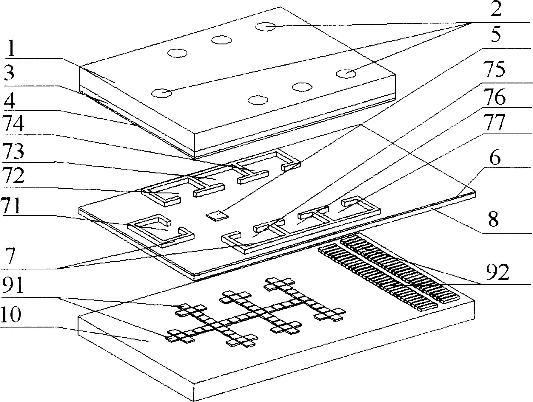
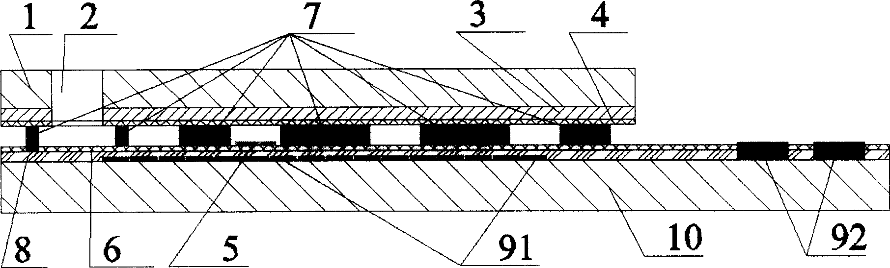
[0052] The lower substrate 10 of this embodiment adopts a silicon wafer material, the control electrode array 91 and the wire 93 adopt a polysilicon material, the lead-out electrode array 92 adopts an aluminum material, the hydrophobic layer 6 uses a Teflon material (Teflon), and the insulating layer 8 adopts silicon dioxide , the support structure 7 is made of SU-8 photoresist, and the detection area 5 (different antibody proteins are used depending on the test object); the material of the upper substrate 1 is glass, and the conductor layer 3 is made of indium thallium oxide (ITO) transparent metal. Teflon is used for the hydrophobic layer 4 on the sheet.

[0053] The manufacturing process of the lower substrate is as follows: firstly, a layer of silicon dioxide is thermally oxidized and grown on the silicon wafer 10, a layer of polysilicon is grown by chemical vap...

Embodiment 2

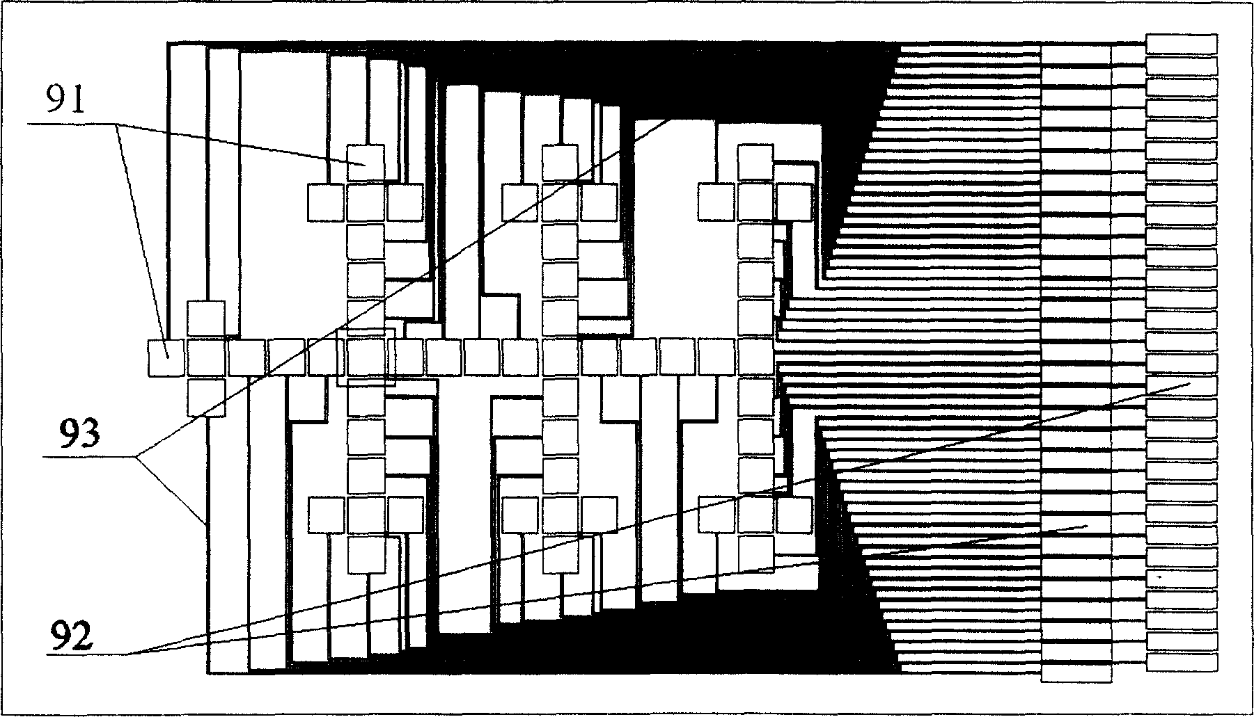
[0067] Example 2 as Figure 5a As shown, the present embodiment is provided with 13 liquid storage chambers, wherein a shared sample chamber 71 is arranged on the top, and the liquid storage chambers are divided into 4 columns (each column has 3 liquid storage chambers) below it, and the control electrode array Distributed between each liquid storage chamber to form a liquid transport channel, three detection areas 51, 52, 53 are arranged side by side on the liquid transport channel, adjacent detection areas 51 and 52 can share the liquid storage chambers 72, 73 and 74, The detection areas 52 and 53 can share the liquid storage chambers 75, 76 and 77, which saves space and reagents. This distribution can simultaneously detect a virus three times in parallel, and the same solid-phase antibody can also be used in the detection areas 51 and 52. and enzyme-labeled antibody, the detection area 53 adopts another solid-phase antibody and enzyme-labeled antibody, so that two kinds of ...

Embodiment 3

[0068] Example 3 as Figure 5b As shown, the present embodiment is provided with 25 liquid storage chambers, wherein the sample chamber 71 is arranged at the central position, and the liquid storage chambers are divided into 8 columns around it (each column has 3 liquid storage chambers), and the control electrode array is distributed in Liquid transport channels are formed between the liquid storage chambers. Four detection areas 51, 52, 53, and 54 are symmetrically distributed in the center of the liquid transport channels, and the distances to each detection area are the same. The solid-phase antibody and The enzyme-labeled antibodies can be different or the same, so that up to 4 viruses can be detected at the same time, or 1 virus can be detected multiple times.


PUM

PropertyMeasurementUnit
Thicknessaaaaaaaaaa

Abstract

A liquid micro drop control chip used in virus detection consists of top and bottom substrates as well as support structure. It is featured as preparing said substrates and said structure by micromechanical means; forming bottom substrate by bottom base plate, electrode array and insulated hydrophobic layer; forming top substrate by top base plate and hydrophobic layer; setting liquid channel, liquid storage region and detection region between top and bottom substrates.

Description

technical field [0001] The invention belongs to the technical field of biochemical detection, and in particular relates to the miniaturization of biochemical detection instruments and the design of micro droplet control devices. Background technique [0002] At present, the qualitative detection of hepatitis virus and HIV can be performed by immunoassays such as blood enzyme-linked immunosorbent assay (Enzyme-Linked Immumosorbnent Assay, ELISA) and immunoassays of its variants for early diagnosis. ELISA is a highly sensitive test technique based on the immunological reaction, which combines the specific reaction of antigen and antibody with the efficient catalytic effect of enzyme on the substrate. Since the reaction of antigen and antibody is carried out in the wells of a polystyrene microtiter plate, which is a solid phase carrier, after each addition of a reagent for incubation, excess free reactants can be removed by washing to ensure the specificity of the test results ...

Claims


Application Information

Patent Timeline
IPC IPC(8): G01N33/569
Inventor 冯焱颖朱亮叶雄英周兆英
Owner TSINGHUA UNIV
Why Eureka
Try Eureka