Wound monitoring sensors and use thereof

a technology for monitoring sensors and wounds, applied in the field of wound monitoring sensors, can solve the problems of traumatic amputation, systemic infection, and increased risk of chronic wounds, and achieve the effects of reducing the capability of continuous monitoring, facilitating stable and selective detection of biomarkers, and reducing the occlusion of biosensors

Active Publication Date: 2021-08-03
UNIV OF MIAMI +1
View PDF32 Cites 0 Cited by
  • Summary
  • Abstract
  • Description
  • Claims
  • Application Information

AI Technical Summary

Problems solved by technology

Monitoring the healing of chronic wounds continues to be a challenge in healthcare.
If the body is unable to heal wounds properly, the patient is at a higher risk of developing localized infections that can lead to systemic infections and traumatic amputations.
Skin ulcers such as diabetic foot ulcers are common wounds that can not only lead to continual breakdown of skin and surrounding tissue, but also increase the risk of deep skin and soft tissue infection.
It is more difficult to recognize and monitor the occurrence of a diabetic wound because of a weakened sensory nervous system that does not give normal pain signals.
The time between the occurrence of a wound and the beginning of treatment can sometimes be lengthy, which can lead to chronic tissue damage.
Even in developed countries, this leads to a more expensive care plan.
Existing wound care sensors are incapable of tracking chronicity from a wound environment.

Method used


Image

  • Wound monitoring sensors and use thereof
  • Wound monitoring sensors and use thereof
  • Wound monitoring sensors and use thereof

Examples


example 1

cterization of Metal Nanostructures

[0146]Platinum, silver and gold nano-material were explored for detection of byproduct, hydrogen peroxide, and UA on the nano-structure modified SPCE and were characterized using SEM (FIG. 6).

[0147]FIG. 6a represents the SEM image of Pt nano-flakes obtained from electro-deposition over a growth period of 15 min at 35° C. This growth period of the flake like structures was optimized over different time periods (15-45 min) and temperatures (35° C.-75° C.). As given in the same SEM images, the Pt nano-flakes have the morphology of thin sheets arranged vertically with numerous edge planes. At 35° C. over 15 min electro-deposition period, the Pt nanoparticles show distinct, regular growth of nano-flakes, with a thickness of platelets in the range of 25 nm to 30 nm with a length of ˜252 nm, at this stage of formation (FIG. 6b). The regularity conformation of these Pt nano-flakes was controlled over different deposition growth periods. SEM characterizatio...

example 2

Enzyme Characterization

[0149]The enzyme UOx was entrapped in PVA-SbQ, a cationic polymer for immobilization on the electrode. The amine group of the styrylpyridinium side chains in the PVA backbone offers firm electrostatic attachment to immobilize the enzyme on the electrode surface. This is due to the net charge of the enzyme being partially negative (6-) above pH 7.5. The pKa and the isoelectric point values of UOx are 4.64 and 7.5, respectively. This binding and interaction of the amino acid chains of UOx with the polymer is depicted in FIGS. 7a and b. The polymer structure in the schematic has minimized bond energy.

[0150]The UOx entrapped in PVA cationic polymer was characterized by various spectroscopic, optical and electrochemical techniques to assess the surface morphology and characteristic behavior. The physisorbed UOx on the electrode surface was used as a control in all these studies. To obtain highest loading of UOx on the electrode surface, various ratios of polymer an...

example 3

Natural Catalyst for UA Detection

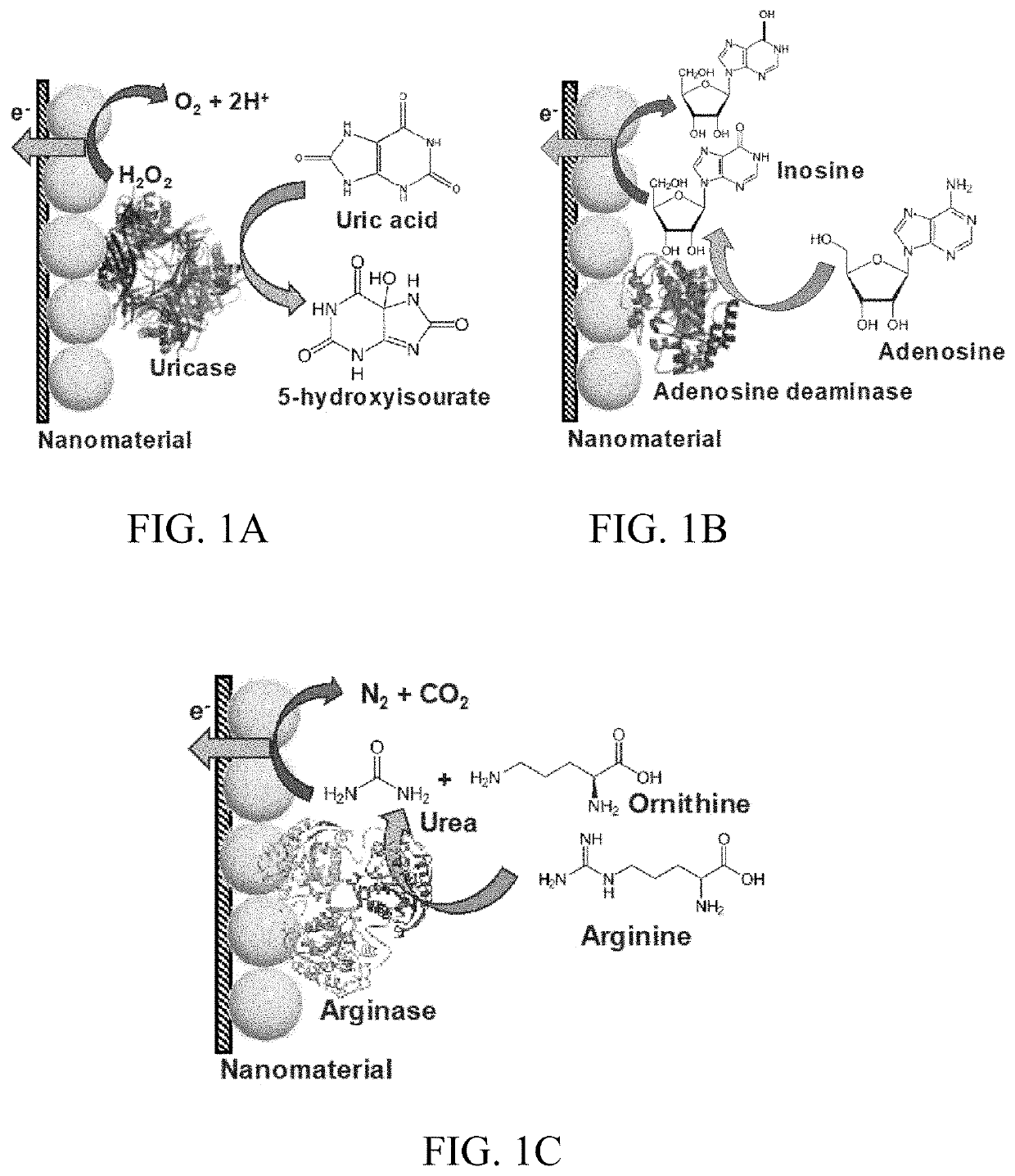
[0158]UOx is an enzyme that belongs to the catabolism of the purine degradation pathway. It plays an integral role in the enzymatic conversion of uric acid (C5H4N4O3) to 5-hydroxyisourate (C5H4N4O4) through oxidation (Eq. 1). The formed 5-hydroxyisourate reacts with water to produce carbon dioxide and allantoin (C4H6N4O3) (soluble form of urate) (Eq. 2). The oxidation of urate to racemic allantoin occurs through the formation of unstable intermediates. In this reaction, C-5 of urate is converted to C-4 of allantoin, while C-2 of urate was recovered as C-2 of allantoin. Yield of the byproduct (H2O2) from this reaction was utilized in well-established colorimetric assay techniques to quantify UA.

UA+O2+H2O→5-hydroxyisourate+H2O2→  (1)

5-hydroxyisourate+H2O→allantoin+CO2→  (2)

[0159]The domain structure of UOx is 50 Å long with a 12 Å wide tunneling fold (T-fold), and is formed from the union of dimers stacked via hydrogen bonds. It is a globular homotetra...


PUM


Abstract

The subject invention provides sensor systems that can detect biomarkers related to wound healing (e.g., uric acid, adenosine, arginine and / or xanthine). In one embodiment, the subject invention pertains to materials and methods for monitoring biomarkers non-invasively in a wound and a biofluid (e.g., sweat) in the proximity of the wound, optionally, including other physiological fluids. Skin based, non-invasive enzymatic electrochemical biosensor on a wearable platform (e.g., sweat patch) that can evaluate the healing of wounds through assessment of its biomarker levels are provided. This non-invasive detection from physiologically biofluids can reduce or eliminate occlusion effects.

Description

CROSS-REFERENCE TO A RELATED APPLICATION[0001]This application claims the benefit of U.S. provisional application Ser. No. 62 / 739,712, filed Oct. 1, 2018, which is incorporated herein by reference in its entirety.STATEMENT OF GOVERNMENT SUPPORT[0002]This invention was made with government support under Grant No. 1160483 awarded by the National Science Foundation Nanosystems Engineering Research Centre for Advanced Self-Powered Systems of Integrated Biosensors and Technologies (ASSIST). The government has certain rights in the invention.BACKGROUND[0003]Monitoring the healing of chronic wounds continues to be a challenge in healthcare. Effective non-invasive approaches could facilitate treatment and reduce the adverse outcomes of trauma, surgery, and amputations associated with chronic wounds.[0004]The wound healing process comprises a series of interdependent physiological events that include: (i) homeostasis, (ii) inflammation, (iii) proliferation, and (iv) maturation. Homeostasis o...

Claims


Application Information

Patent Timeline
Patent Type & Authority Patents(United States)
IPC IPC(8): A61B5/1486A61B5/20A61B5/145A61B5/00
CPCA61B5/1486A61B5/14507A61B5/14539A61B5/14546A61B5/20A61B5/4266A61B5/4277A61B5/14517A61B5/445A61B5/6833A61B2560/0412
Inventor CHOUDHURY, SOHINI ROYUMASANKAR, YOGESWARANBHANSALI, SHEKHARKIRSNER, ROBERT S.LEV-TOV, HADAR A.
Owner UNIV OF MIAMI
Why Eureka
Try Eureka