Three dimensional corneal imaging with gabor-domain optical coherence microscopy

a gabor-domain optical coherence and gabordomain technology, applied in the field of three-dimensional corneal imaging with gabor-domain optical coherence microscopy, can solve the problems of insufficient accuracy of specular microscopy, poor regenerative capacity of endothelial cells in vivo, and introduce subjectivity and error. , to achieve the effect of enhancing accuracy and reducing bias in cell quantification

Active Publication Date: 2021-08-17
LIGHTOPTECH CORP
View PDF5 Cites 0 Cited by
  • Summary
  • Abstract
  • Description
  • Claims
  • Application Information

AI Technical Summary

Benefits of technology

[0011]The present disclosure is directed to enhancing the accuracy of and reducing bias in cell quantification and particularly endothelial cell density (ECD) quantification by providing Gabor-domain optical coherence microscopy (GDOCM) for three dimensional (3D) wide field of view (1 mm2) corneal imaging and machine learning for automatic delineation of endothelial cell boundaries.
[0013]Generally, the present disclosure provides a method of imaging a tissue sample, including the steps of imaging, by contact imaging or non-contact imaging, a portion of the tissue sample to create image data; applying a low pass filter to the image data to create a first filtered data; thresholding the first filtered data to create thresholded data; median filtering the thresholded data to reduce thresholding artifacts; automatically identifying target cell structures through iterative machine processing to reduce outliers to define a fitted surface; and adjusting an intensity projection of a slice adjacent the fitted surface to accommodate errors introduced by at least one of (i) the image thresholding and (ii) median filtering to provide substantially artifact free en face view of the portion of the tissue sample.
[0014]In one configuration, an apparatus is provided for imaging a tissue sample, the apparatus including a Gabor-domain optical coherence microscope (GDOCM) configured for imaging, by contact imaging or non-contact imaging, of a portion of the tissue sample; and a processor connected to the GDOCM, the processor configured to (i) apply a low pass filter to the image data to create a first filtered data; (ii) threshold the first filtered data to create thresholded data; (iii) median filter the thresholded data to reduce thresholding artifacts; (iv) automatically identify target structures through iterative machine processing to reduce outliers to define a fitted surface; and (v) adjust an intensity projection of a slice adjacent the fitted surface to accommodate errors introduced by at least one of (aa) the image thresholding and (bb) median filtering to provide a substantially artifact free en face view of the portion of the tissue.
[0015]The present disclosure further contemplates a method of assessing tissue graft efficacy, including the steps of in vivo imaging, by contact imaging or non-contact imaging, a grafted tissue with a Gabor-domain optical coherence microscope; applying a low pass filter to the image data to create a first filtered data; thresholding the first filtered data to create thresholded data; median filtering the thresholded data to reduce thresholding artifacts; automatically identifying target cells through iterative machine processing to reduce outliers to define a fitted surface; and adjusting an intensity projection of a slice adjacent the fitted surface to accommodate errors introduced by at least one of (aa) the image thresholding and (bb) median filtering to provide a substantially artifact free en face view of the grafter tissue.

Problems solved by technology

Optical coherence tomography (OCT) has been shown to be comparable to ultrasound, while specular microscopy has been determined to be insufficiently accurate.
These limits from specular microscopy may cause under-sampling.
Subsequently, the actual cell counting is typically performed manually, which introduces an associated subjectivity and error.
Further, endothelial cells show poor regenerative capacity in vivo and are critical for preserving corneal transparency vis-a-vis their fluid pumping mechanism that maintains appropriate hydration status and clarity of the stroma.
This immersion further leads to inaccuracies.
Prior methods lack sufficient robustness to deal with images of varying quality, as they are prone to under- and over-segmentation in regions with poor image quality.

Method used


Image

  • Three dimensional corneal imaging with gabor-domain optical coherence microscopy
  • Three dimensional corneal imaging with gabor-domain optical coherence microscopy
  • Three dimensional corneal imaging with gabor-domain optical coherence microscopy

Examples


Embodiment Construction

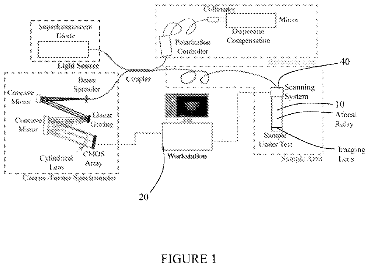
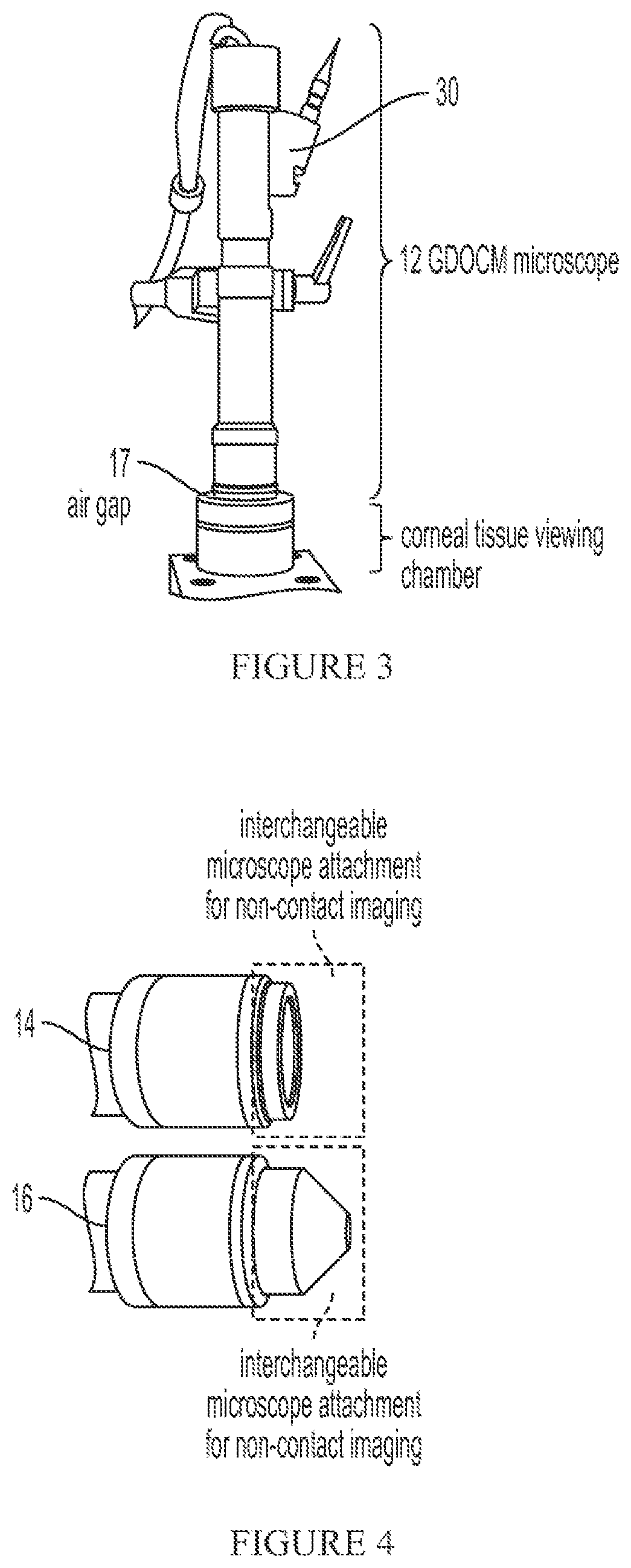
[0038]The present system provides for non-contact imaging of tissue, such as corneal tissue, through a viewing chamber with Gabor-domain optical coherence microscopy (GDOCM) over a larger field-of-view than specular microscopy (SM). A three dimensional (3D) image processing algorithm is applied to segment the endothelium and correct the corneal curvature (nonplanar orientation of the cells), resulting in artifact-free images of the flattened endothelium.

[0039]Although the present description is set forth in terms of tissue that includes corneal endothelial cells, it is understood the tissue can includes retinal cell mosaics, such as photoreceptors, retinal pigment epithelium (RPE) cells, or any other suitable cells. Further, it is understood that images of the tissue may be acquired ex vivo, in vivo, or in vitro.

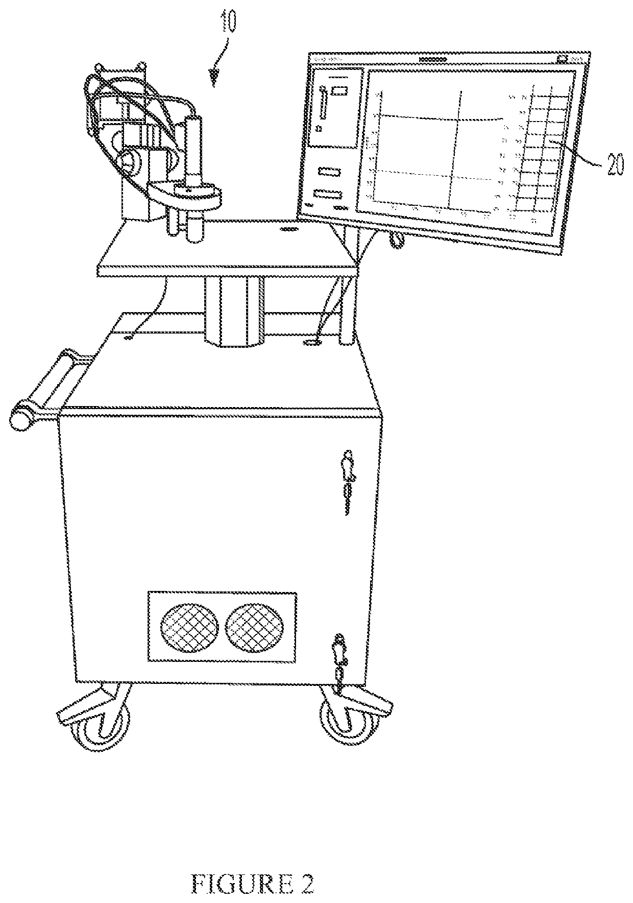
[0040]Referring to FIG. 1, the present system includes a microscope 10 for generating image data and a workstation or controller 20 for processing the acquired image data. I...


PUM


Abstract

A system for non-contact imaging of corneal tissue stored in a viewing chamber using Gabor-domain optical coherence microscopy (GDOCM), wherein a 3D numerical flattening procedure is applied to the image data to produce an at least substantially artifact-free en face view of the endothelium.

Description

CROSS-REFERENCE TO RELATED APPLICATIONS[0001]This application claims the benefit of U.S. Provisional patent application 62 / 822,406 filed Mar. 22, 2019 entitled OPTICAL AND NUMERICAL METHODS FOR MICROSCOPIC TISSUE ANALYSIS.STATEMENT REGARDING FEDERALLY SPONSORED RESEARCH OR DEVELOPMENT[0002]This invention was made with government support under Grant Number: 1R43EY028827-01, awarded by NIH National Eye Institute, and Grant No. IIP-1534701, awarded by the National Science Foundation. The government has certain rights in the invention.THE NAMES OF THE PARTIES TO A JOINT RESEARCH AGREEMENT[0003]Not applicable.REFERENCE TO A SEQUENCE LISTING[0004]Not applicable.STATEMENT REGARDING PRIOR DISCLOSURES BY THE INVENTOR OR A JOINT INVENTOR[0005]Not applicable.BACKGROUND OF THE INVENTION[0006]Eye banks typically conduct three main exams to qualify of corneal tissue: (i) evaluation of stromal opacity; (ii) pachymetry; and (iii) assessment of endothelial cell density and morphology.[0007]The evalu...

Claims


Application Information

Patent Timeline
Patent Type & Authority Patents(United States)
IPC IPC(8): G06K9/00G06T7/00G06T11/00
CPCG06T7/0014G06T11/008G06T2207/10028G06T2207/20032G06T2207/20081G06T2207/30041G06T2207/30024G06T7/12G06T7/136G06T5/002G06T2207/10056G06T2211/424G06V20/64G06V40/193G06V10/30G06V10/454G06V10/82G06V10/764G06T2211/456
Inventor CANAVESI, CRISTINACOGLIATI, ANDREA
Owner LIGHTOPTECH CORP
Why Eureka
Try Eureka