Joint estimation diffusion imaging (JEDI)

a diffusion imaging and joint estimation technology, applied in the field of joint estimation diffusion imaging, can solve the problems of limiting the ability of reliable estimation, limiting the utility of both research and clinical applications, and prone to errors in the standard art of both methods, and achieve the effect of convenient implementation

Active Publication Date: 2021-09-28
RGT UNIV OF CALIFORNIA
View PDF50 Cites 0 Cited by
  • Summary
  • Abstract
  • Description
  • Claims
  • Application Information

AI Technical Summary

Benefits of technology

[0012]In one embodiment, a new method is provided for combining standard single pulsed field gradient (sPFG) diffusion weighted magnetic resonance (dMRI) data used for estimation of local macroscopic diffusion anisotropy and global tractography with double pulsed field gradient (dPFG) dMRI data used for estimation of local microscopic diffusion anisotropy into a single estimation scheme that provides more accurate estimates of both local diffusion anisotropy and global connectivity. This method combines previously-disclosed methods for morphological shape analysis and segmentation (SWD), for simultaneous local diffusion estimation and global tractography (GO-ESP), and a new method called Diffusion Tensor Subspace Imaging (DiTSI) for dPFG analysis, as well as a fast and accurate approach for non-linear registration between modalities (SYMREG), combined using the joint estimation procedure for integrating multi-modal data (JESTER). This joint analysis method is capable of extracting new levels of information that is not achievable from sPFG or dPFG modalities alone. The new theoretical dPFG analysis (DiTSI), combined with a new theoretical framework for joint sPFG+dPFG analysis (JEDI) within the probabilistic framework of JESTER allow construction of quantitative diffusion anisotropy and connectivity measures previously unattainable.

Problems solved by technology

Nevertheless, the problem of tissue heterogeneity continues to plague current standard dMRI methods, limiting its ability to provide reliable estimates of both local and global tissue properties, thus ultimately limiting its utility in both research and clinical applications.
Standard art in both methods is error prone due to complications with tissue fiber heterogeneity.
However, as has been discussed in the literature, sPFG methods are ultimately limited in the presence of voxels whose internal tissue structure is so complex as to render their macroscopic anisotropy negligible, with gray matter (GM) voxels being the most important example.
In fact, analytical models for more realistic tissue microstructures would be difficult to obtain, and alternative approaches are needed.

Method used


Image

  • Joint estimation diffusion imaging (JEDI)
  • Joint estimation diffusion imaging (JEDI)
  • Joint estimation diffusion imaging (JEDI)

Examples


example 1

xel Simulation

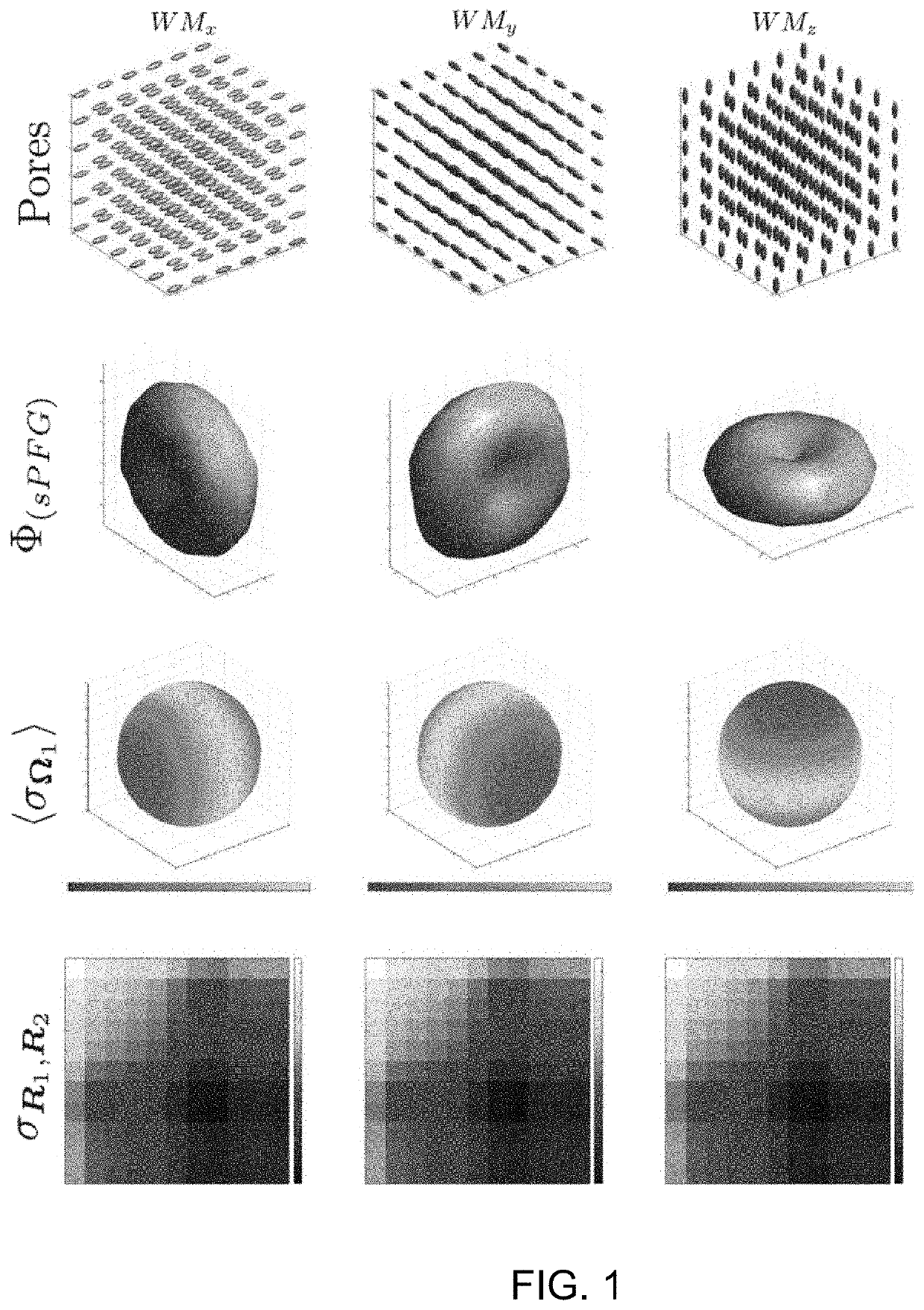
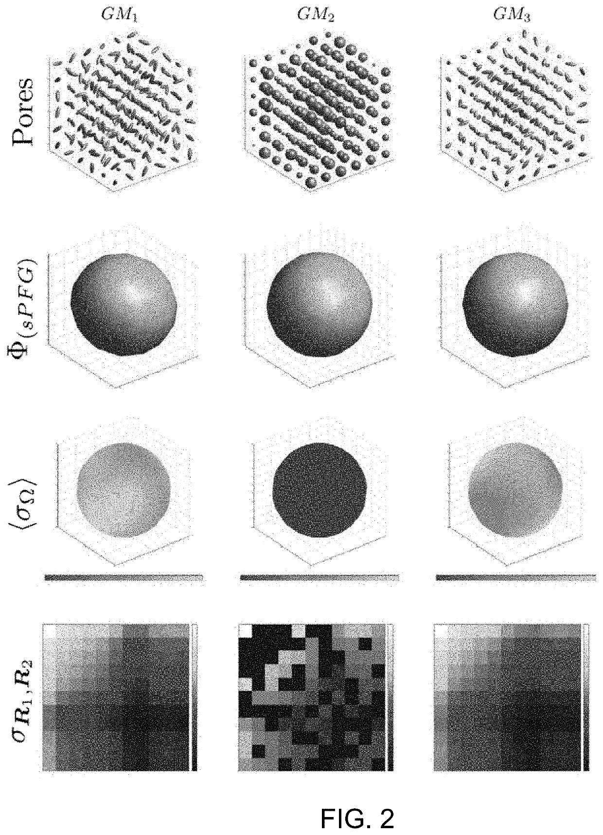
[0080]An illustrative example of DiTSI is provided by simulating the signal from a single voxel with structures composed of idealized WM and GM geometries. The voxel diffusion characteristics were constructed in a similar fashion as disclosed by Özarslan, et al., J. Chem Phys. 2009; 130:104702 (incorporated herein by reference) by assuming that the voxel is discretized into p equal sections in each direction and thus p3 cubic subvolumes (or “pores”), each of which contained a simple ellipsoidal diffusion profile characterized by a real symmetric 3×3 diffusion tensor characterized by the three principal eigenvectors and their associated eigenvalues {λ1, λ2, λ3}. The simulation input allowed arbitrary manipulation of the relative eigenstructure of these ellipsoids, facilitating the generation of various “tissue” models. For illustrative purposes, mitigation of angular sampling discretization provides more informative examples and so a large number of unique diffusion ang...

example 2

on of DiTSI to Human dPFG Data

[0085]Acquisition and reconstruction parameters: To assess the translation of DiTSI into real human applications, we developed a modified version of the Extended Hybrid-space SENSE for EPI (EHSS-EPI,) pulse sequence able to collect sPFG and dPFG encoding in both the original dual spin echo (dPFG-DSE) and the single spin echo (dPFG-SSE) implementation. This method uses an off-resonance and eddy current corrected joint interleaved blip-up / down reconstruction and provides an efficient method for fast and accurate (i.e., distortion-free) acquisition, which will be critical for practical applications. Initial tests revealed that the dPFG-SSE version had significantly better signal-to-noise than the dPFG-DSE version and thus was the method used in the human studies. All data were collected on a General Electric (GE) Discovery MR750 3.0T Whole Body clinical imaging system using a 32-channel from NOVA Medical Devices (Wilmington, Mass.).

[0086]The dPFG sampling ...

example 3

on of JEDI to Human dPFG and sPFG Data

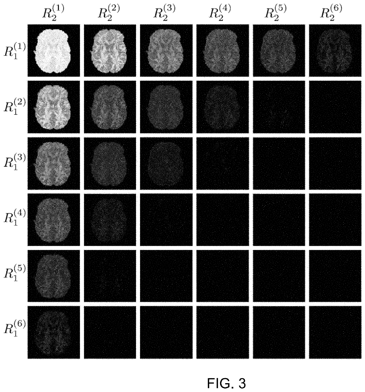
[0089]Acquisition and reconstruction parameters: To test the JEDI method, sPFG diffusion acquisition data was combined with the dPFG data acquired using the same EHSS-EPI pulse sequence (and reconstruction) described above. The protocol acquires 3 shells at b-values of b=(1000, 2000, 3000 s / mm2 with the number of directions being 30, 45, and 60, respectively. The data acquisition parameters included slice thickness thk=2 mm, FOV=20 cm, TE=119 ms, TR=4.0 s, matrix size=100×100×72 with a SENSE factor of 4. The imaging time was 16 min. A standard high resolution anatomical (HRA) dataset was also collected using an inversion recovery prepared MP-RAGE protocol, 1.2 mm sagittal slices (S / I direction), Locs / slab=170, number of slabs=1, FOV=26 cm, TR / TE / FA=7 ms / 3 ms / 8°, Inversion time TI=900 ms, matrix size 256×256, BW=31.25 kHz, NEX=1.

[0090]Results: The JEDI method, as an extension of the GO-ESP method, produces the same output: local anisotropy define...


PUM


Abstract

A method and for estimating local diffusion anisotropy and global tractography within neural architecture from diffusion weighted magnetic resonance image (dMRI) data uses a computer processor to integrate a first dataset comprising standard single pulsed field gradient (sPFG) dMRI data with a second dataset comprising double pulsed field gradient (dPFG) dMRI data into a common coordinate system with the same spatial resolution. The resulting image includes integrated macroscopic and microscopic anisotropy and global tractography within the target volume.

Description

RELATED APPLICATIONS[0001]This application claims the benefit of the priority of U.S. Provisional Application No. 62 / 857,185, filed Jun. 4, 2019, which is incorporated herein by reference in its entiretyGOVERNMENT RIGHTS[0002]This invention was made with government support under Grant No. MH096100 awarded by the National Institutes of Health and Grant No. DBI1147260 awarded by the National Science Foundation. The government has certain rights in the invention.BACKGROUND[0003]The sensitivity of diffusion weighted MR imaging (dMRI) methods to tissue microstructure continues to tantalize the research and clinical community with its promise of quantitative characterization of both local tissue microstructure through the analysis of intravoxel diffusion anisotropy and global tissue connectivity via the reconstruction of intervoxel fiber pathways (i.e., tractography). Nevertheless, the problem of tissue heterogeneity continues to plague current standard dMRI methods, limiting its ability ...

Claims


Application Information

Patent Timeline
Patent Type & Authority Patents(United States)
IPC IPC(8): G01R33/563A61B5/055G01R33/56
CPCG01R33/56341A61B5/055G01R33/5608A61B5/0042
Inventor FRANK, LAWRENCE R.GALINSKY, VITALY L.
Owner RGT UNIV OF CALIFORNIA
Why Eureka
Try Eureka