
Method to measure the activation state of signaling pathways in cells

a signaling pathway and activation state technology, applied in the field of biological and chemical analysis of molecules in cells, can solve the problems of inability to detect the activity of one or more additional proteins within the cell, and inability to detect the activity of one or more additional proteins in the cell

Inactive Publication Date: 2002-09-12
RGT UNIV OF CALIFORNIA
View PDF5 Cites 53 Cited by
  • Summary
  • Abstract
  • Description
  • Claims
  • Application Information

AI Technical Summary

Benefits of technology

0023] The methods described above can be used for any purpose in combination with two dimensional gel electrophoresis, protein mass spectroscopy, yeast 2-hybrid assays, structural biology, intracellular ion and other indicators, intracellular protein location techniques, DNA arrays, and flow cytometry (including sheathed and unsheathed flow and flow cytometry on a microfluidics device). The protein activity of three or more proteins is detected and/or quantified and preferably the protein activity of four, five, six, ten or more proteins is detect

Problems solved by technology

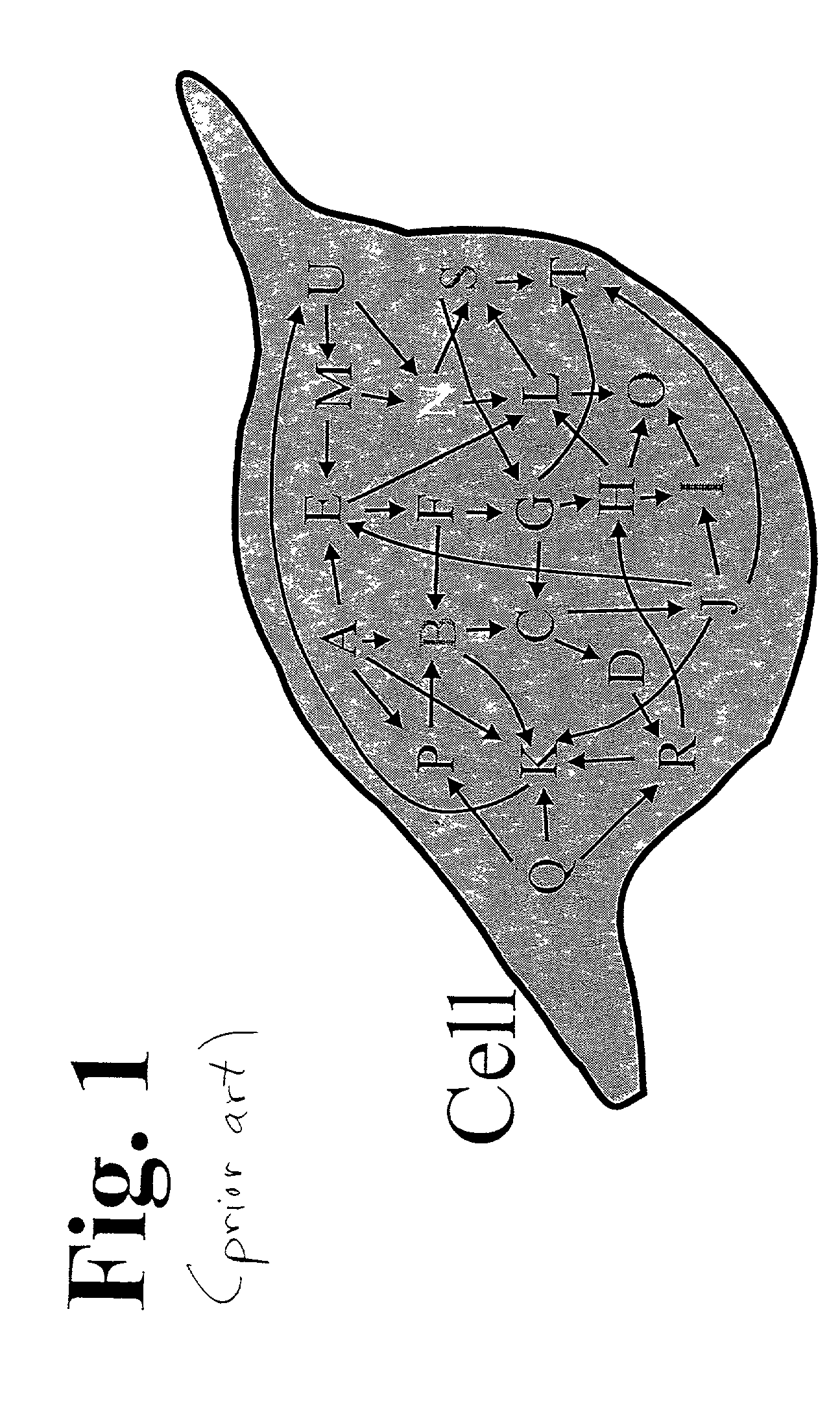
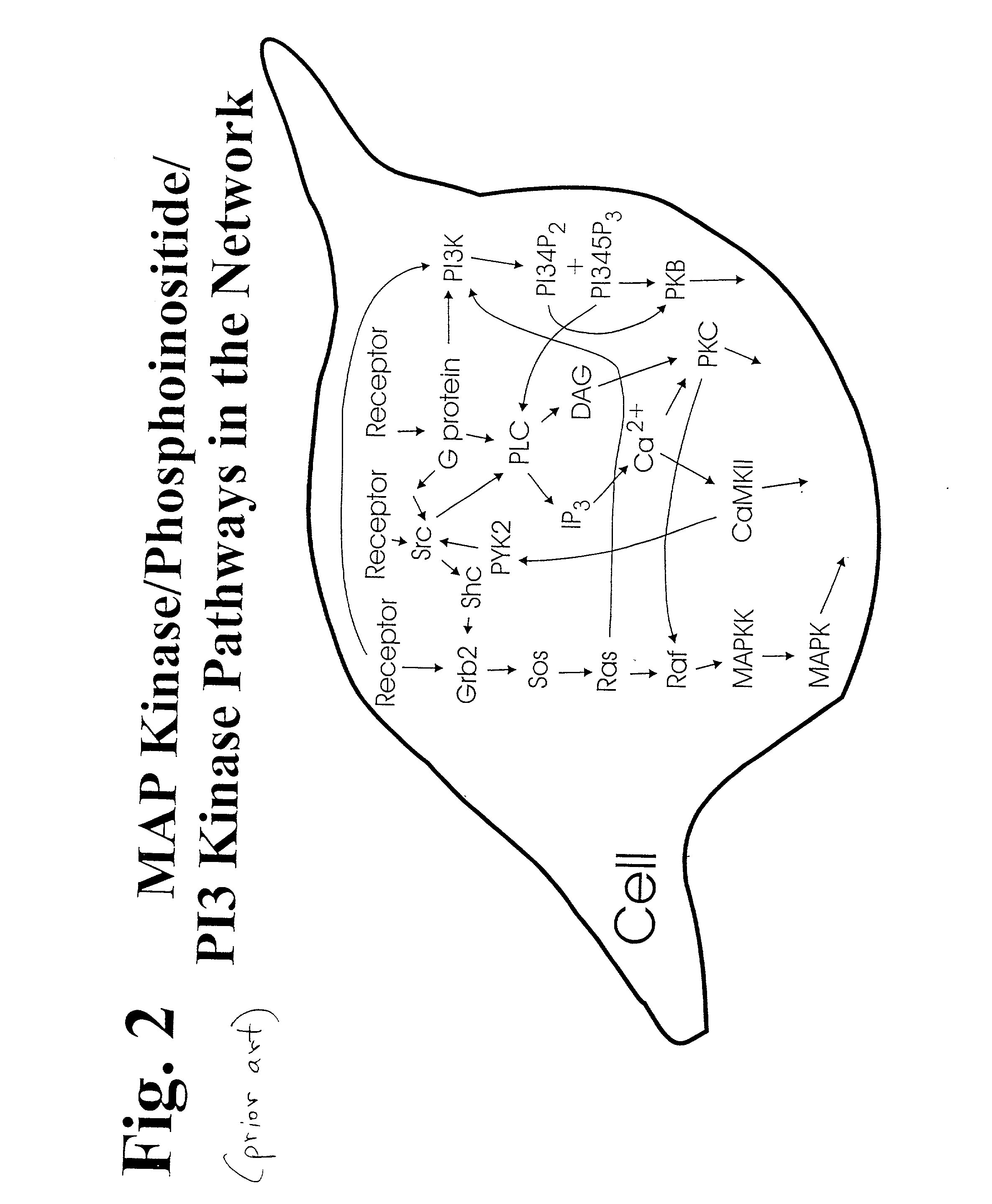
The determination of which proteins are active in a living cell and how networks of proteins interact in a living cell is a complex problem in biology and in pharmaceutical development.
Disease often results from a breakdown in communication between and within cells.
A challenge in developing drugs against kinases and other types of enzymes is to find molecules that are specific for only the enzyme of interest.
Many potential drugs may have a desired effect on its expected target, but may also have an undesired effect on the activity of one or more additional proteins within the cell.
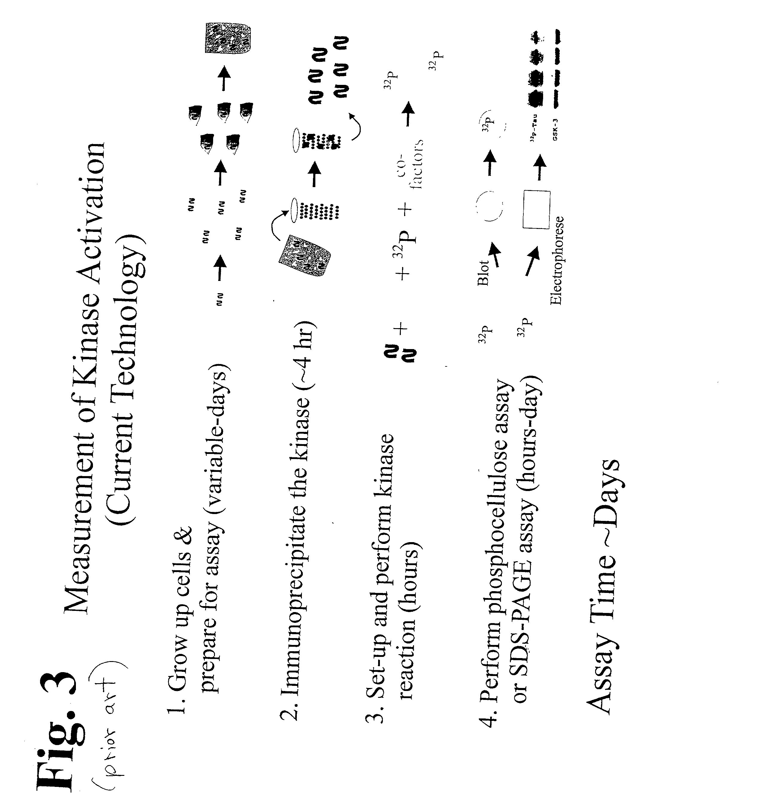
Presently available in vitro biochemical assays for enzyme activity often give misleading results since other enzymes and modulators of enzyme activity present in a living cell are not present in the assay (Table I).
These factors are difficult to add to an assay because their identity and concentration are often unknown.
The situation is further complicated by the fact that the concentration of these factors and the activity of the proteins can change over time (Table II).
Additionally, since the proteins exist in interconnected networks, the activities of many proteins will be affected by perturbations in the cellular environment or by the abnormal activity of another protein within the networks.
This inappropriate activation leads to the uncontrolled growth of blood cells that is manifested as cancer.
However, the presence of the gene or its protein product is not perfectly correlated with the appearance of cancer.
Existing techniques for the measurement of enzyme activation in a single cell, group of cells, or a portion of a cell have inherent limitations.
Unfortunately, current proteomic technologies have significant shortcomings in the study of enzyme function.
This technique gives a very high resolution for protein separations, but it is difficult to perform, and it cannot detect many important cellular proteins, especially those in low abundance or those with a hydrophobic character (traits typical of many if not most enzymes).
The new mass spectrometry techniques combined with bioinformatics have improved the identification of proteins separated by electrophoresis techniques, but do not solve the fundamental issues of performance difficulty and sensitivity.
The new chip-based methods hold the as yet unfulfilled promise for identifying large numbers of proteins quickly; however, sensitivity, specificity, and quantification are still issues to resolve.
However, these approaches do not directly measure the critical parameter of the most crucial proteins: the activity of enzymes.
Attempts are made to measure a protein's activity by the identification of a phosphorylated species (many proteins are "switched on" by the addition of one or more phosphate groups), but such an approach can provide only an indirect measure of activity, and detecting such species in a single cell, group of cells, or a portion of a cell is fraught with difficulty.
There are three major drawbacks to this approach: 1) the methods have poor sensitivity so that the cytoplasm of large numbers of cells must be pooled; 2) a time-averaged level of the kinase activity is actually measured since the cells are not synchronous with respect to their activation status; and 3) the normal relationships of the pathways and networks within which signal transduction enzymes reside are disrupted.
Similar drawbacks exist for measurement of the activity of most other types of enzymes.
Unfortunately, both inactive and active enzyme molecules are highlighted by this method, although some attempt may be made to infer the activity level of the enzyme from its location within the cell.
This strategy has worked well for the measurement of various ion concentrations (i.e., Ca.sup.2+), but thus far has not been generally applicable to the measurement of enzyme activity in the living cell.
A further limitation to all of these techniques is their inability to measure multiple enzyme activities simultaneously in a cell.

Method used


Image

  • Method to measure the activation state of signaling pathways in cells
  • Method to measure the activation state of signaling pathways in cells
  • Method to measure the activation state of signaling pathways in cells

Examples


Embodiment Construction

[0061] The activity of multiple proteins in a single living cell, portion of a cell or in a group of cells are simultaneously examined. A database is compiled from the application of this method to cells under a large variety of different conditions. The protein activity is measured by introducing one or more reporter molecules (substrates) into one or more cells. The reporter(s) is chemically modified by the enzyme of interest. In some cases the enzyme(s) of interest is affected by the addition of a stimulus or a pharmaceutical compound to the cell. The reactions between the enzymes and the reporters are diminished or terminated, and the reporter and modified reporter are removed from the cell. The activity of the enzyme(s) is determined by measuring the amount of reporter molecules remaining, by measuring the amount of altered reporter molecules produced, or by comparing the amount of reporter molecules to the amount of altered reporter molecules. A database is compiled of the act...


PUM


Abstract

The activity of multiple proteins in a single living cell, portion of a cell or in a group of cells is simultaneously measured by introducing reporter molecules. The reporter(s) is chemically modified by the enzyme of interest. In some cases the enzyme(s) is affected by the addition of a stimulus or a pharmaceutical compound to the cell. The reactions between the enzymes and the reporters are diminished or terminated, and the reporter and modified reporter are removed. The activity of the enzyme(s) is determined by measuring the amount of reporter remaining, the amount of altered reporter produced, or by comparing the amount of reporter to the amount of altered reporter. A database is compiled of the activities of the different proteins. By performing a series of experiments at different time points, conditions, and varieties of cell types, a database is developed for molecular cellular mechanisms in health and disease states. By exposing cells to a variety of compounds data for drug development and screening is provided.

Description

RELATED APPLICATIONS[0001] This application is related to and claims priority from U.S. Provisional Patent Application Ser. No. 60 / 252,861, filed Nov. 22, 2000, and is a continuation-in-part application of application Ser. No. 09 / 358,505 filed Jul. 21, 1999 and entitled "Method and Apparatus for Detecting Enzymatic Activity Using Molecules that Change Electrophoretic Mobility", which was in turn a continuation-in-part application of application Ser. No. 09 / 036,706 filed Mar. 6, 1998, and entitled "Fast Controllable Laser Lysis of Cells for Analysis" now issued as U.S. Pat. No. 6,156,576 (2000).BACKGROUND OF THE INVENTION[0002] 1 Field of the Invention[0003] This invention relates to the areas of the biochemical and chemical analysis of molecules in cells, and in particular to an assay and method for measuring the activation of internal chemical activity of a plurality of proteins in a single cell, a population of cells, or portion of a cell.[0004] 2. Description of the Prior Art[000...

Claims


Application Information

Patent Timeline
IPC IPC(8): B01L3/00G01N1/02G01N15/14G01N33/50G01N33/68
CPCB01L3/5027G01N1/02G01N15/1475G01N33/5005G01N33/6803G01N15/1433
Inventor ALLBRITTON, NANCYSIMS, CHRISTOPHER
Owner RGT UNIV OF CALIFORNIA
Learn More