AHF associated dispersion system and method for preparation

a dispersion system and associated technology, applied in the field of ahf associated dispersion system and method for preparation, can solve the problems of increasing the cost of patient discomfort, irreversible loss of protein structure, and inability to meet the needs of patients, and achieves the effects of reducing the intensity of peaks, increasing temperature, and reducing the ellipticity at 295 nm

Inactive Publication Date: 2002-09-19
THE RES FOUND OF STATE UNIV OF NEW YORK +1
View PDF8 Cites 21 Cited by
  • Summary
  • Abstract
  • Description
  • Claims
  • Application Information

AI Technical Summary

Benefits of technology

0057] Temperature induced changes in rhAHF secondary structure was studied by acquiring the far-UV CD spectra (255 nm to 205 nm, FIG. 1A). At 20.degree. C., a broad negative band at 215 nm suggested that the protein existed predominantly in .beta.-sheet conformation. This is in agreement with the structure, proposed based on homology modeling (Pan, Y. T., DeFay, T. et al. (1995) "Proposed structure of the A domains of factor VIII by homology modelling [letter]." Nat Struct Biol 2(0): 740-4, which is hereby incorporated by reference in its entirety). As the temperature is increased over the temperature range of from 20.degree. C. to 50.degree. C., there were no significant changes in the far-UV CD spectrum; indicating that the secondary structure of the protein was not altered. In the temperature range of from 50.degree. C. to 65.degree. C., the ellipticity at 215 nm increased progressively with increasing temperature suggesting an increase in the .beta.-sheet conformation. At temperatures over 65.degree. C., significant changes were observed in the spectral characteristics. The CD spectra had red-shifted by approximately 2 nm and appearance of a positive band in the range from 205 nm to 210 nm range suggested the formation of anti-parallel .beta.-strands possibly leading to formation of aggregates eventually stabilized by intermolecular .beta.-strands (Hilbich, C., Kisters-Woike, B. et al. (1991) "Aggregation and secondary structure of synthetic amyloid beta A4 peptides of Alzheimer's disease." J Mol Biol 218(1): 149-63; Hammarstrom, P. M., Persson, M. et al. (1999) "Structural mapping of an aggregation nucleation site in a molten globule intermediate." J Biol Chem 274(46): 32897-903, which are hereby incorporated by reference in their entirety). Thus the secondary structure appeared to undergo the following conformational transition:
0058] Far-UV spectral data was used to calculate the F.sub.app, the apparent fraction in the unfolded form, according to the method described herein.
0059] Analysis of the profile indicated that the melting of the protein appeared to occur in two distinct stages. The first transition was sharp and prominent, occurring approximately at the range of from 60.degree. C. to 62.degree. C. and may possibly be associated with the melting of heavy chain of the protein while the second transition observed at about 70.degree. C., was relatively broad. Since rhAHF is a multi-domain protein the observed multi-stage transition may be attributed to the unfolding of heavy and light chains of the rhAHF at different temperatures.
0060] While the near-UV CD spectrum is indicative of the tertiary structure, the far-UV CD spectrum is indicative of the secondary structure. We investigated the temperature dependence of the near-UV CD spectrum over the wavelength range of from 255 nm to 320 nm, (FIG. 1B). At 20.degree. C., there were two positive peaks at about 295 nm and about 268 nm and as the temperature was increased, the intensity of the peaks decreased.
0061] The near-UV CD spectrum was used to calculate the temperature dependence of the unfolding of tertiary structure. With increase in temperature, ellipticity at 295 nm decreased and thus F.sub.app increased with midpoint of main transition occurring approximately in the range of from 50.degree. C. to 52.degree. C., (FIG. 1C). The temperature dependence of far-UV CD spectrum was monitored over the wavelength range of from 205 nm to 255 nm and the main transition detected by far-UV CD was considerably higher, approximately from 60.degree. C. to 62.degree. C., (FIG. 1C). Such a difference in the temperature at which tertiary and secondary structural changes occurred confirmed the existence of intermediate unfolded state(s). (Ptitsyn, O. B., Pain, R. H. et al. (1990) "Evidence for a molten globule state as a general intermediate in protein folding." FEBS Lett 262(1): 20-4, which is hereby incorporated by reference in its entirety). While the multistage transition was apparent for the far-UV CD spectra, it was relatively less apparent for the near-UV CD spectra.
0062] Factor VIII is a multi-domain protein with several tryptophan residues and changes in Trp fluorescence may provide information on gross tertiary structural changes in the protein, spanning different domains. Fluorescence emission spectra of rhAHF were acquired over the temperature range of from 25.degree. C. to 90.degree. C. to detect changes in Trp fluorescence, (FIG. 2). The data indicated multistage transitions and they did not overlap with the one observed by far-UV CD. The midpoint of the main transition was approximately in a range of from 50.degree. C. to 52.degree. C. and was almost similar to the one observed for the near-UV CD spectrum. Over the temperature range of from 20.degree. C. to 50.degree. C., there was an approximately 33% decrease in the intensity of the emission at 330 nm. Over the same temperature range of from 20.degree. C. to 50.degree. C., the far-UV CD data indicated that the secondary structure of the protein was intact suggesting the existence of a structured intermediate (SI.sub.1) in the unfolding pathway. Between from 50.degree. C. to 65.degree. C., it was observed that the decrease in fluorescence intensity was approximately 44% and this temperature range was also associated with the main transition occurring at about 52.degree. C. (near-UV CD data) indicating significant loss of tertiary structure. The far-UV CD data also showed a transition over this temperature range and indicated a corresponding increase in the .beta.-sheet conformation suggesting the existence of the second structured intermediate (SI.sub.2). Between from 65.degree. C. to 90.degree. C., a further 23% decrease in intensity was observed and the fluorescence profile indicated complete loss of tertiary structure. The far-UV CD studies above 65.degree. C. indicated that the protein had a tendency to form intermolecular .beta.-strands suggesting the existence of the third structure intermediate (SI.sub.3). Thus, our initial analysis showed that there were three distinct population of intermediate structures in the unfolding pathway; SI.sub.1 with unaltered secondary structure and small changes in the tertiary structure; SI.sub.2 with significant loss of tertiary structure and increased .beta.-sheet conformation and SI.sub.3 with complete loss of tertiary structure and anti-parallel .beta.-strands.

Problems solved by technology

However, the commercially available protein pharmaceutical has been reported to undergo the aforementioned physical instability problems with concomitant loss of therapeutic activity (Baxter Health Care, unpublished results), thus requiring a new formulation strategy.
This translates into not only higher cost but also patient discomfort.
In the presence of liposomes, while the formation of SI.sub.1 was completely reversible, appearance of SI.sub.2 resulted in irreversible loss of protein structure.

Method used


Image

  • AHF associated dispersion system and method for preparation
  • AHF associated dispersion system and method for preparation
  • AHF associated dispersion system and method for preparation

Examples


example 1

Preparation of Liposomes

Conventional Method

[0047] In the procedure according to the conventional method: 0.30 mg / ml DMPC, 0.15 mg / ml bPS and 0.04 mg / ml DSPE-PEG dissolved in chloroform were taken in a round-bottomed flask and the solvent was removed using a rotary evaporator, depositing the lipid as a thin film along the walls of the flask. Multilamellar vesicles (MLV) encapsulating the protein were formed by dispersing the lipid film in the appropriate buffer (0.4 M NaCl and 50 Mm Tris) containing 0.5 mg / ml of the protein, with gentle swirling at room temperature.

Present Methodology

Method 1

[0048] In the procedure according to the present method: 0.30 mg / ml DMPC, 0.15 mg / ml bPS and 0.04 mg / ml DSPE-PEG dissolved in chloroform were taken in a round-bottomed flask and the solvent was removed using a rotary evaporator, depositing the lipid as a thin film along the walls of the flask. Multilamellar vesicles (MLV) encapsulating the protein were formed by dispersing the lipid film in the a...

example 2

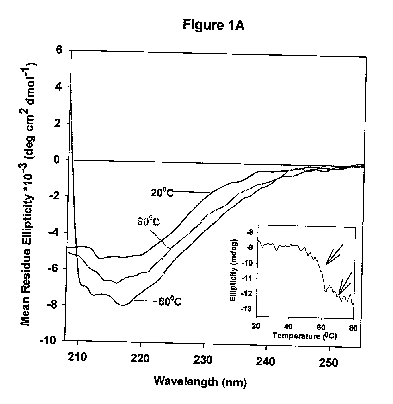
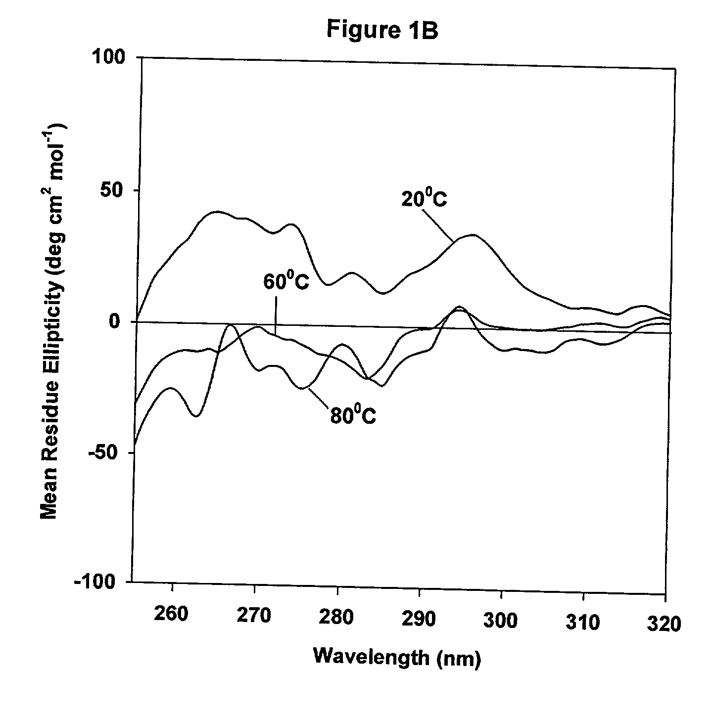
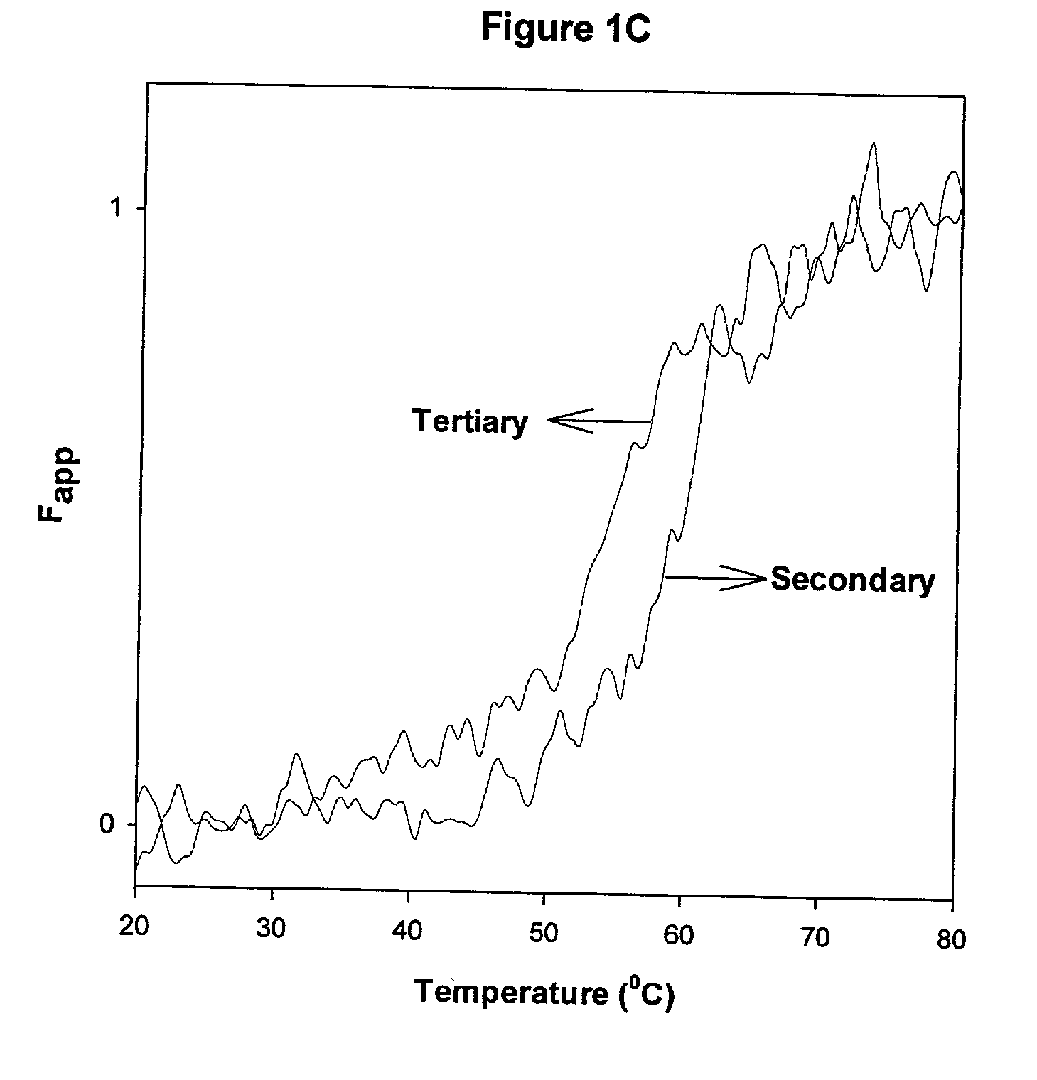
Circular Dichroism Experiments

[0050] CD spectra were acquired on a JASCO-715 spectropolarimeter calibrated with d10 camphor sulfonic acid. Samples were scanned in the range of from 205 nm to 255 nm for secondary structure analysis, and typically, the protein concentration used was about from 20 .mu.g / ml to 22 .mu.g / ml. For near-UV CD studies, spectra were acquired in the range of from 320 nm to 255 nm, using a 10 mm quartz cuvette, and the protein concentration used was about 0.5 mg / ml. CD spectra of the protein were corrected by subtracting the spectrum of the buffer baseline and multiple scans were acquired and averaged to improve signal quality. The CD spectra of samples containing liposomes may be distorted as a result of light scattering. The contribution due to light scattering was corrected as follows: (1) the ellipticity values at from 350 nm to 400 nm were monitored and used as a baseline that was subtracted from the scans; (2) multiple scans were acquired and averaged to i...

example 3

Fluorescence Studies

[0051] Fluorescence spectra were acquired on a SLM 8000C spectrofluorometer (Urbana, Ill.). The intensity of the emission spectra was monitored over the range of from 300 nm to 400 nm, using a slit width of 4 nm on the excitation and emission paths. The excitation monochromator was set at 280 nm and a 295 nm long pass filter was used to minimize scattering effects. The melting of the protein was followed by monitoring the decrease in the intensity of the emission at 330 nm over the temperature range of from 25.degree. C. to 90.degree. C. Samples were equilibrated at the desired temperature for approximately 3 to 4 minutes using a water bath (Neslab RTE 110).


PUM

PropertyMeasurementUnit
temperatureaaaaaaaaaa

Abstract

A method for complexing AHF in a dispersed medium, includes: a) providing an AHF protein, b) altering the conformational state of the AHF protein to expose hydrophobic domains therein, c) binding a stabilizer to the exposed hydrophobic domains, and d) at least partially reversing the alteration to associate at least a portion of the protein with the stabilizer. A pharmaceutically effective stabilized AHF dosage wherein above about 0.5%, preferably above about 3%, and more preferably above about 25%, of the AHF molecule is associated with a stabilizer is also disclosed.

Description

[0001] This application claims the benefit of U.S. Provisional Patent Application Serial No. 60 / 250,137, filed Nov. 30, 2000 (which is hereby incorporated by reference in its entirety).[0002] The present invention relates to Antihemophilic ("AHF") protein associated with a dispersion system and methods for preparation thereof. In particular, the present invention relates to liposomal association, including encapsulation, of AHF proteins and methods for preparation thereof.DESCRIPTION OF THE RELATED ART[0003] Hemophilia A is an inherited bleeding disorder caused by deficiency of antihemophilic factor, a blood coagulation protein that functions as a cofactor in the coagulation cascade (Lamer, A. J. (1987) "The molecular pathology of haemophilia." Q J Med 63(242): 473-91, which is hereby incorporated by reference in its entirety). It is transmitted as a defect on the X chromosome and affects 1 male in 5,000. The severity of the bleeding disorder varies among patients depending upon the...

Claims


Application Information

Patent Timeline
Patent Type & Authority Applications(United States)
IPC IPC(8): A61K9/127A61K38/37A61K47/10A61K47/18
CPCA61K9/1271A61K38/37A61K47/10A61K47/18
Inventor BALASUBRAMANIAN, SATHYAMANGALAM V.BESMAN, MARCKASHI, RAMESHRAMANI, KARTHIK
Owner THE RES FOUND OF STATE UNIV OF NEW YORK
Why Eureka
Try Eureka