Cardiovascular imaging and functional analysis system

a functional analysis and cardiac imaging technology, applied in the field of medical diagnostic and screening apparatuses and methods, can solve the problems of crystals, position-based, and pixellated scintillation, and the improvement of detector hardwar

Inactive Publication Date: 2002-12-12
NORTH COAST IND INC
View PDF0 Cites 81 Cited by
  • Summary
  • Abstract
  • Description
  • Claims
  • Application Information

AI Technical Summary

Problems solved by technology

However, medical instrument improvements have not kept pace with detector hardware enhancements such as pixellated scintillation crystals, and position-sensitive photomultipliers.
The single crystal Anger camera has numerous disadvantages when used for first-pass RNA related to its low count rate capability including pulse-pileup and low count density.
A low-count density image limits the ability to define changes in wall motion.
This scenario provides for improved count rate capability but limits spatial resolution.
In practice, clinical interpretations of the SIM400 scanner treadmill stress acquisitions are more difficult to analyze.
Patient motion during peak stress acquisition can cause image artifacts that can trigger an incorrect diagnosis.
Unfortunately, this method only corrects for up / down / side planar motion (i.e., motion in a plane, and motion in any direction outside the defined plane is uncorrected).
However, the prior art instruments were slow, designed primarily for relatively low rate (up to 5 kHz) breast imagers.
Also, its rate capability is in fact limited by the physical nature of radiation interaction with a gas detector medium in a two-step process.
Neither of these strategies has produced a solution to the high incidence of previously asymptomatic CAD deaths.
In summary, nuclear cardiology equipment has evolved in recent years in the direction of more complex and expensive devices.
The risk of transporting these patients to nuclear medicine departments inhibits currently optimal utilization of these diagnostic techniques.
In this form, the test is much less stressful and can be performed on patients for whom a standard stress test would be difficult to perform or even dangerous.
Prior art gamma cameras in medical practice have intrinsic rate capability limited to less than .about.100 kHz due in part to their slow front-end electronics and data acquisition systems.
The small-size multicrystal camera developed by Picker International, now Marconi, can operate at high rates but has poor spatial resolution, has very limited software capabilities and is very expensive.
It is not possible to obtain in valve location in equilibrium pool studies.
In principle, a single head system equipped with a special high energy collimator can be used in positron imaging, but this result comes at the expense of reduced spatial resolution and sensitivity.
For example, an ultrasound examination can last up to 30 minutes and will not provide all useful information such as pulmonary transit time, as compared to a 15 second duration of the first pass test providing this information.
For example in women attenuation of 140 keV photons in radiodense breasts produce attenuation artifacts in heart imaging, potentially obscuring blood flow deficiencies.
Other competing or potentially competing technologies for dedicated heart imagers, such as CdZnTe solid state detectors (developed by Siemens, GE Medical), CsI(Tl) scintillator / silicon photodiodes (Digirad), and gas based detectors (Proportional Technologies) are not capable of efficiently imaging positron emitters.
GSO and LSO scintillators are faster and have higher stopping power for 511 keV photons than NaI(Tl) but are much more expensive and have lower energy resolution (resulting in lower scatter rejection) for lower energy photons, such as 140 keV from Tc-99m.

Method used


Image

  • Cardiovascular imaging and functional analysis system
  • Cardiovascular imaging and functional analysis system
  • Cardiovascular imaging and functional analysis system

Examples


Embodiment Construction

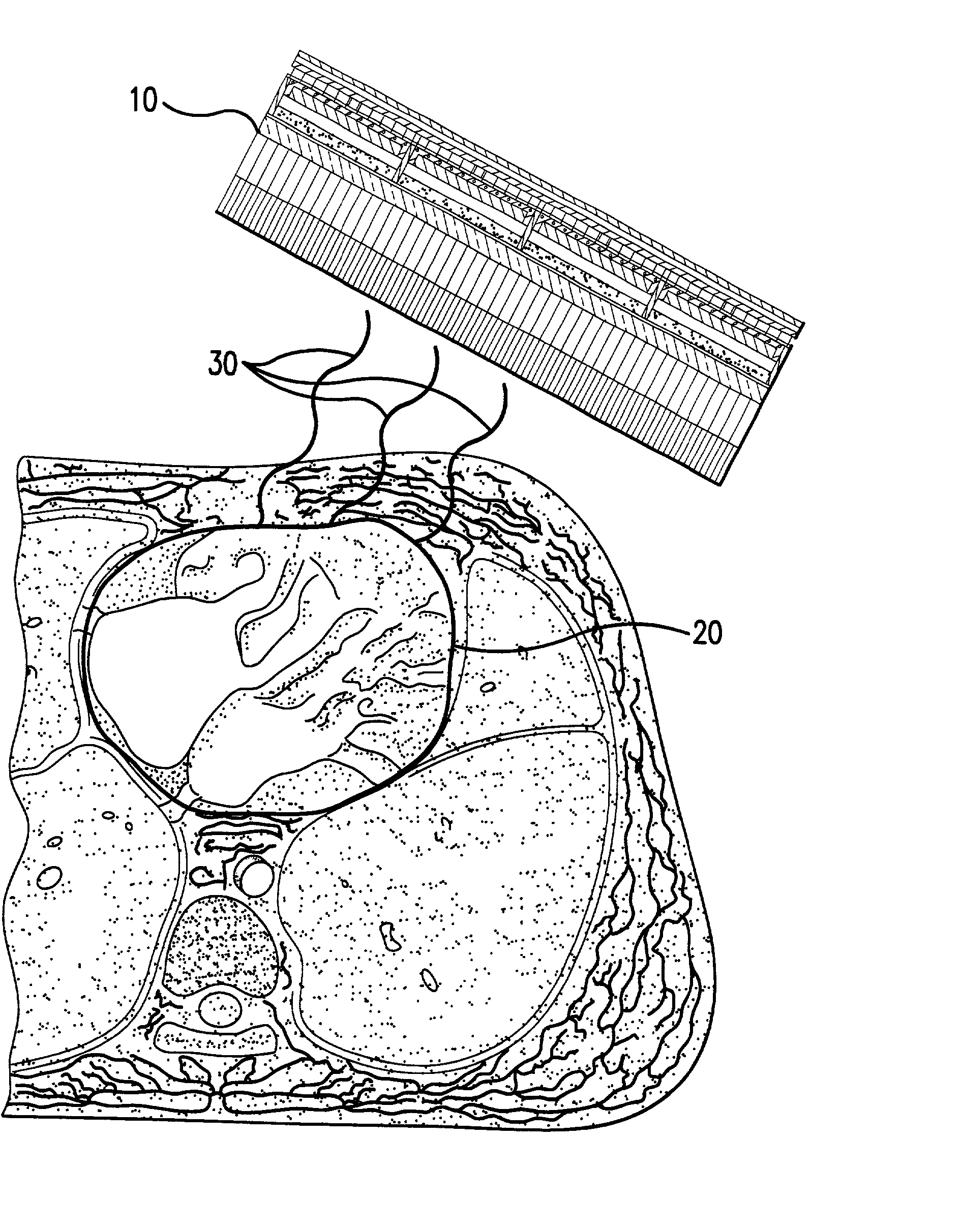
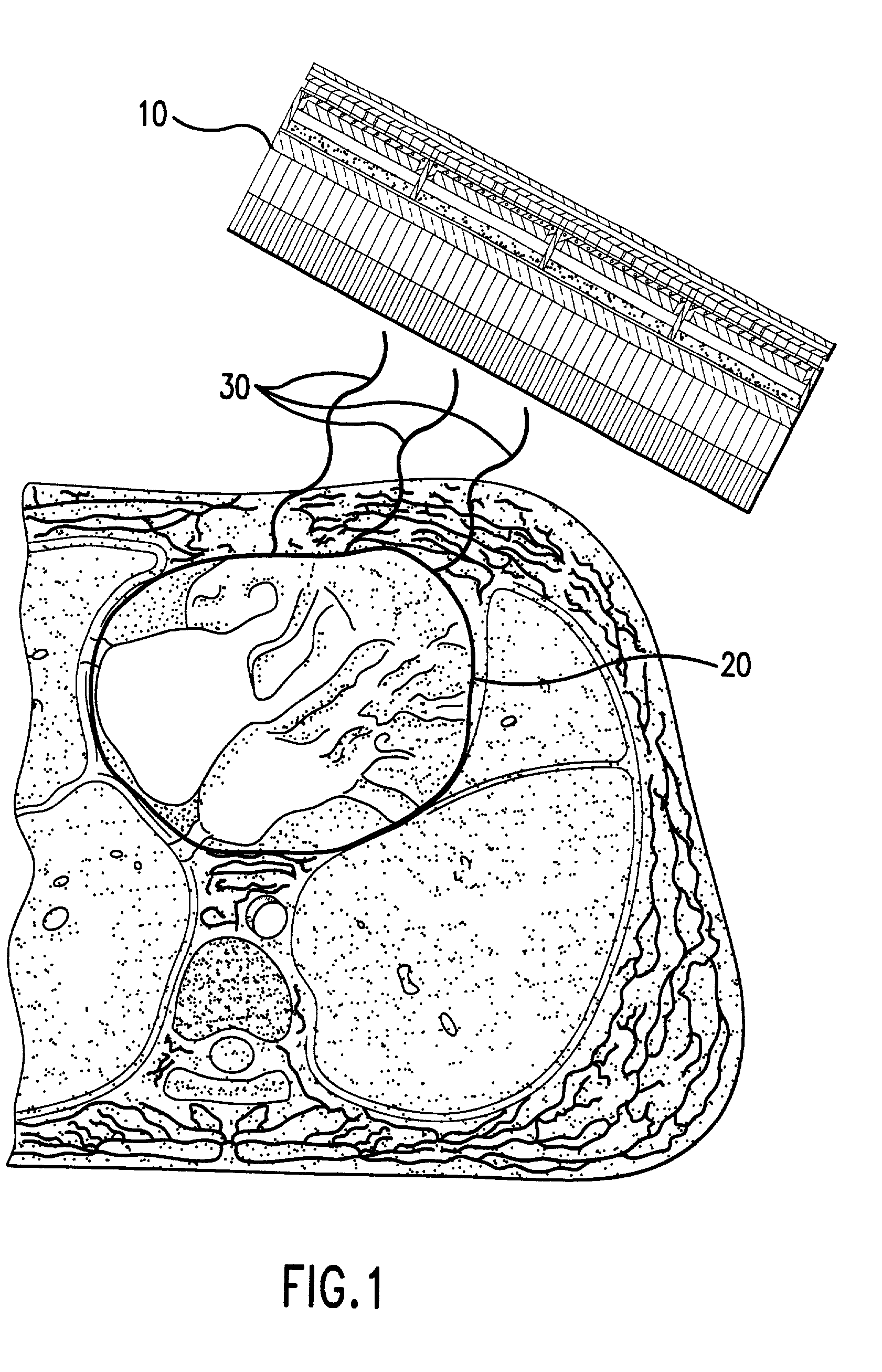
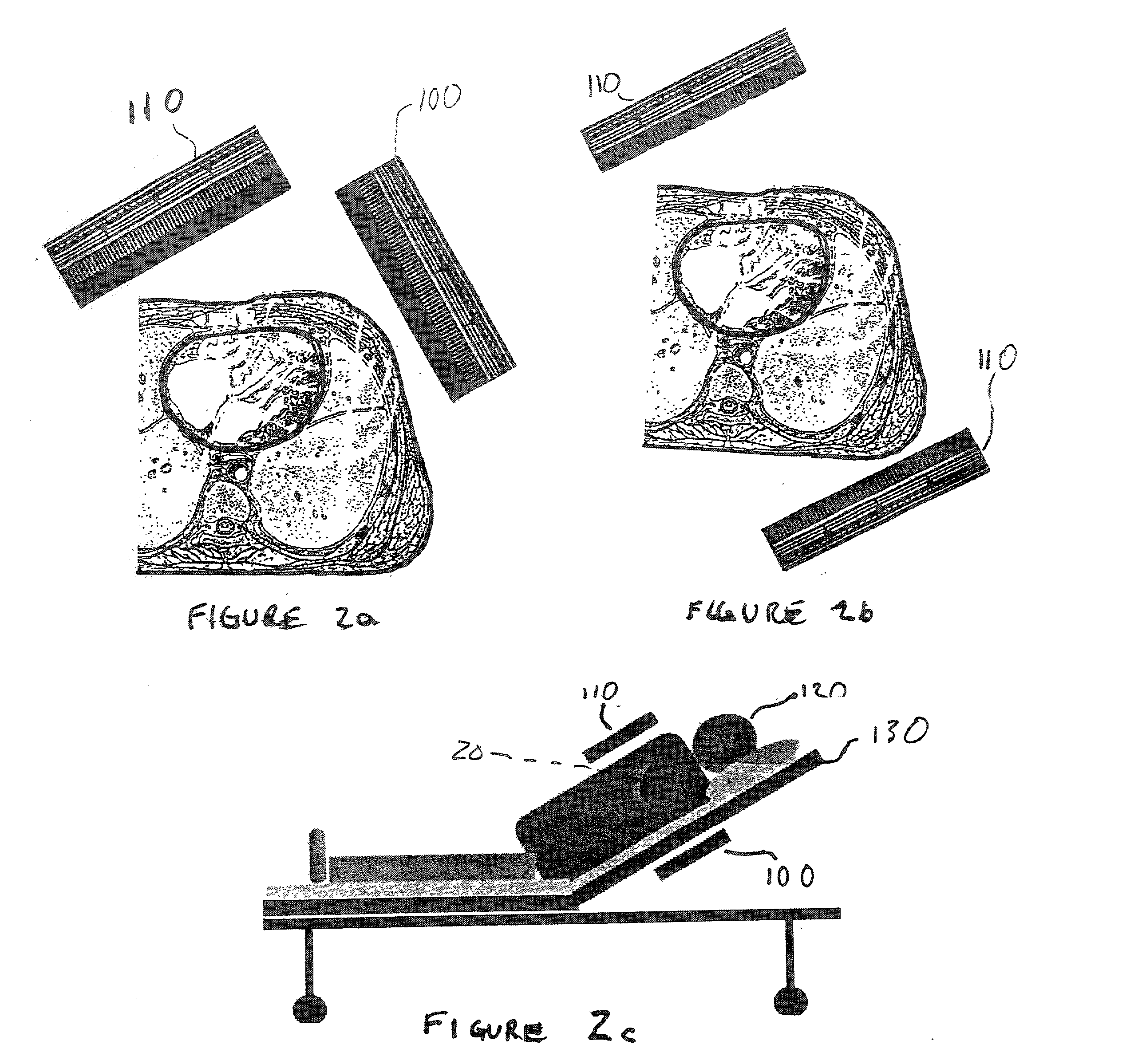
[0096] To enhance further the understanding of the cardiovascular imaging and functional analysis system disclosed herein, the following example of a specific embodiment of the present invention is presented. As disclosed, the example presented is a Cardiovascular Non-Invasive Screening Probe System and Method for Coronary Artery Disease.

[0097] The components of the exemplary Non-Invasive Screening Probe System include the following:

[0098] Two dedicated gamma probes

[0099] Fast signal processing electronics and fast data acquisition system

[0100] Gantry for control electronics and computer

[0101] Support arm(s) for the detector probes

[0102] Computer system with data processing algorithm

[0103] Digital data storage system

[0104] Hardcopy printer

[0105] The exemplary Non-Invasive Screening Probe System, in accordance with an embodiment of the present invention provides an economical instrument to ascertain coronary artery disease. During the screening procedure, the patient is injected intr...


PUM


Abstract

A Cardiovascular imaging and functional analysis system and method employing a dedicated fast, sensitive, compact and economical imaging gamma camera system that is especially suited for heart imaging and functional analysis. The system uses a dedicated nuclear cardiology small field of view imaging camera, allowing image physiology, while offering inexpensive and portable hardware. In some variations, a basic modular design suitable for cardiac imaging with one of several radionucleide tracers is used. The detector is positioned in close proximity to the chest and heart from several different projections, allowing rapid accumulation of data for first-pass analysis, positron imaging, quantitative stress perfusion, and multi-gated equilibrium pooled blood tests. In one variation, a Cardiovascular Non-Invasive Screening Probe system provides rapid, inexpensive preliminary indication of coronary occlusive disease by measuring the activity of emitted particles from an injected bolus of radioactive tracer.

Description

[0001] 1. Field of the Invention[0002] The present invention relates to medical diagnostic and screening apparatuses and methods. Particularly, the present invention relates to non-invasive medical diagnostic and screening systems. Still more particularly, the present invention relates to non-invasive imaging and functional analysis systems for evaluating cardiac and cardiovascular health.[0003] 2. Related Art[0004] Cardiac imaging and functional analysis is the largest single nuclear medical imaging application and represents the area of greatest unmet need in the prior art. This need is exemplified by the fact that historically, for 30%-50% of those stricken with coronary occlusive (artery) disease, the first symptom of the disease is death. This outcome has motivated considerable interest in developing diagnostic methods and apparatuses to detect the condition of coronary occlusive disease prior to the onset of fatal symptoms, and further to assist in the development and implemen...

Claims


Application Information

Patent Timeline
Patent Type & Authority Applications(United States)
IPC IPC(8): A61B5/0275
CPCA61B5/02755A61B6/4258A61B6/507
Inventor BISHOP, HARRYMAJEWSKI, STANISLAWUMENO, MARC M.
Owner NORTH COAST IND INC
Why Eureka
Try Eureka