
Novel assay for detecting immune responses involving antigen specific cytokine and/or antigen specific cytokine secreting T-cells

Inactive Publication Date: 2003-07-31
THE GOVERNMENT OF THE UNITED STATES OF AMERICA AS REPRESENTED BY THE SEC OF THE NAVY NAVAL RES LAB WASHINGTON
View PDF0 Cites 11 Cited by
  • Summary
  • Abstract
  • Description
  • Claims
  • Application Information

AI Technical Summary

Benefits of technology

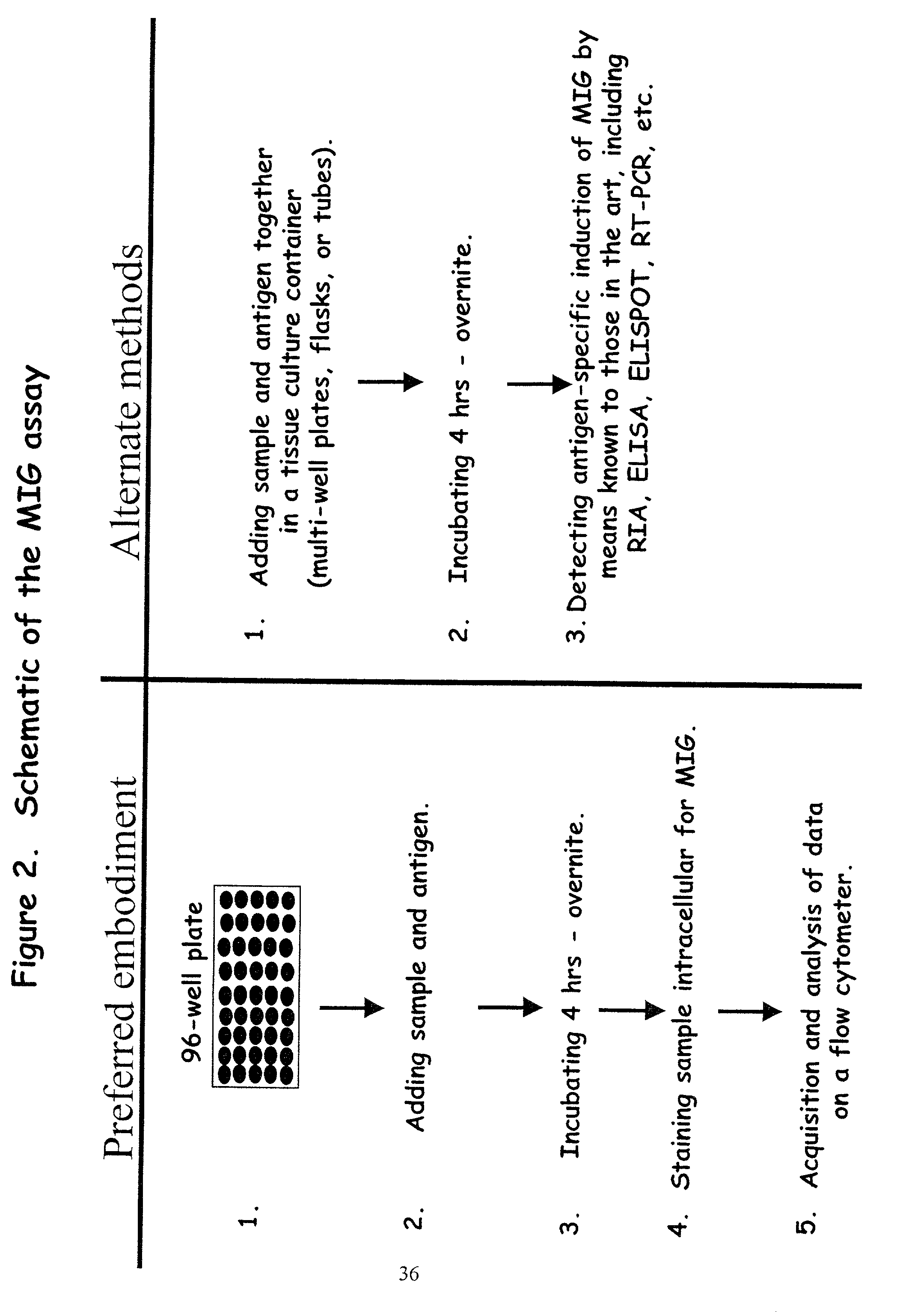
0045] In addition to sensitivity, the instant assay has a number of additional advantages over other assays. For example, antigen-specific IFN-.gamma. restricted responses can be readily detectable by MIG assay following a few hours of incubation with antigen. In the examples provided herein, antigen-specific induction of MIG expression was detectable following 4-16 hours of culture. In contrast, those of skill in the art frequently utilize longer culture time for the ELISPOT assay (24-36 hours and as long as 13 days). Additionally, even if the variable culture time is discounted from each method the MIG assay is at least 2.85 times faster to perform from start to finish than the ELISPOT assay which is comparable to other current detection methods. (Table IV). Thus, the MIG assay also provides a method for detecting antigen specific IFN-.gamma. responses that is less laborious and requires less time to complete and evaluate.
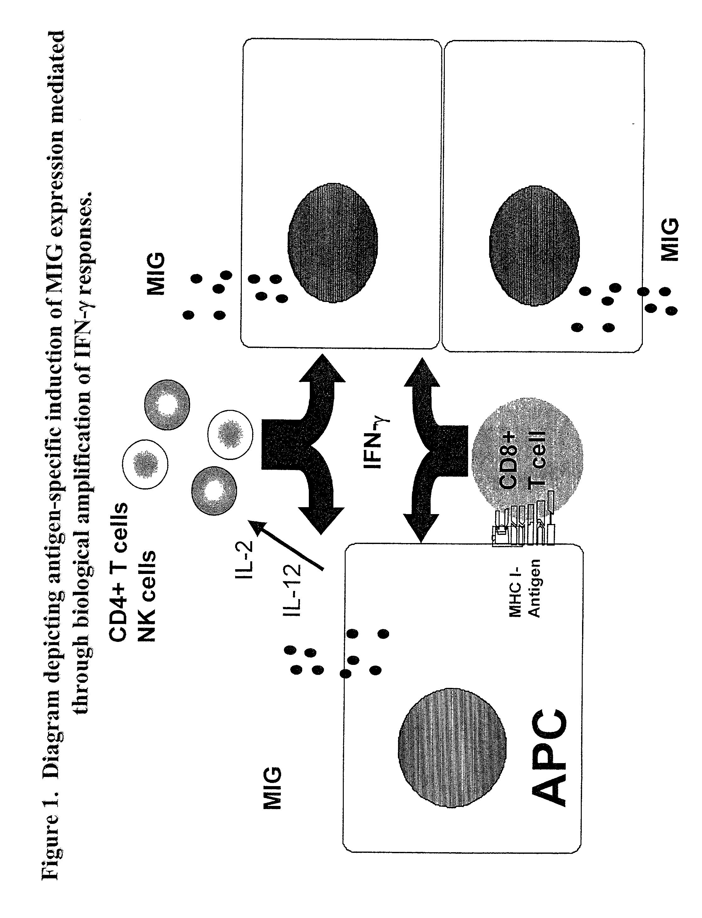
0046] Furthermore, because the assay of the present invention can be a flow cytometry-based assay, it can be technically simple and a relatively rapid and objective method for identifying antigen-specific cells. However, current protocols for detecting antigen-specific cytokine producing cells by intracellular staining and flow cytometry do not provide a robust means of reproducibly detecting low levels of antigen-specific, cytokine producing cells at significant levels above background. Because antigen-specific induction of MIG expression is the result of biological amplification of IFN-.gamma. responses, it provides a novel alternative for detecting low levels of antigen-specific, IFN-.gamma. restricted responses by flow cytometry and a measure of immune responsiveness. Detection of MIG expression by flow cytometry evaluates the frequency of responsive cells and magnitude of response at a large-scale, single cell level, as compared with the ELISPOT assay where it is the number of responsive cells in a limited sample in each well that are detected and enumerated. Antigen-specific detection of MIG expression by flow cytometry is more sensitive than the IFN-.gamma. ELISPOT assay because it is based on the biological amplification of IFN-.gamma. responses, allowing the detection of positive responses that are not scored positive by the IFN-.gamma. ELISPOT assay conducted in parallel. As shown in the Examples disclosed herein, the use of CD8.sup.+ T cell depletion and add-back experiments provides one example demonstrating that the MIG assay can be at least two times more sensitive than the IFN-.gamma. ELISPOT assay.
0047] In a typical embodiment, the invention uses a new assay for detecting IFN-.gamma. other Th-1 cytokines or cells secreting IFN-.gamma. or other Th-1 cytokines by measuring one of the biological effects of the activity of the cytokine through flow cytometric detection of the chemokine whose producti

Problems solved by technology

To date, the detection of low levels of antigen-specific cellular immune responses has been problematic, particularly in human systems.
However, these conventional assays are cumbersome and laborious, require extensive tissue culture and are not sensitive enough to detect low frequencies of antigen-specific cells.
Furthermore, they can not be used to evaluate responses associated with cells which may respond to specific antigen by cytokine production, for example, but may not proliferate.
However, prolonged in vitro cultivation and variations of culture conditions may not accurately reflect in vivo immunologically relevant events.
Additionally, the laboratory procedure for completion of the ELISPOT assay is time consuming.
Furthermore, quantitation of ELISPOT is achieved by subjective manual counting or by computerized counting microscopes which provide an objective and reproducible means of enumerating ELISPOTs, however, the cost of these machines do not make them readily available to most laboratories.
Moreover, ELISPOT assays require additional time for coating plates and typically longer culture times and an additional day for development for the assay.
Tetramer staining is more sensitive than traditional methods for detecting antigen-specific cells, but the use of this technique is restricted to well characterized epitopes in association with defined MHC alleles, and requires subsequent culture for functional characterization of tetramer-positive cells.
Flow-based intracellular staining methods provide a technically simple and relatively fast method for identifying antigen-responsive cells; however, it is difficult to detect low numbers of antigen-specific cytokine-producing cells at levels significantly above background.
The limitation of the assay lies in the fact that low frequency of cytokine producing cells may be indistinguishable above background levels.
Addition of immune enhancer reagents, such as antibodies to CD28, are frequently used to augment costimulation in culture conditions but these reagents may bias the results.
Very limited success has been reported for the detection of ex vivo antigen-specific cells in CD4.sup.+ or CD8.sup.+ T cells from immunized individuals where the number of circulating antigen-specific cells may be considerably lower than that found in individuals exposed to the infectious agent.
Indeed, without using tetramers to select a subpopulation of specific cells for evaluation, it has not been possible to detect low frequencies of antigen-specific cells using flow cytometry for intracellular cytokines.
IG. This results in an inability of IFN-.ga
Moreover, there is no description of how MIG expression could be used as a marker for detecting antigen-specific cells or for detecting antigen-specific IFN-.gamma. producing cells.

Method used


Image

  • Novel assay for detecting immune responses involving antigen specific cytokine and/or antigen specific cytokine secreting T-cells
  • Novel assay for detecting immune responses involving antigen specific cytokine and/or antigen specific cytokine secreting T-cells
  • Novel assay for detecting immune responses involving antigen specific cytokine and/or antigen specific cytokine secreting T-cells

Examples


example 2

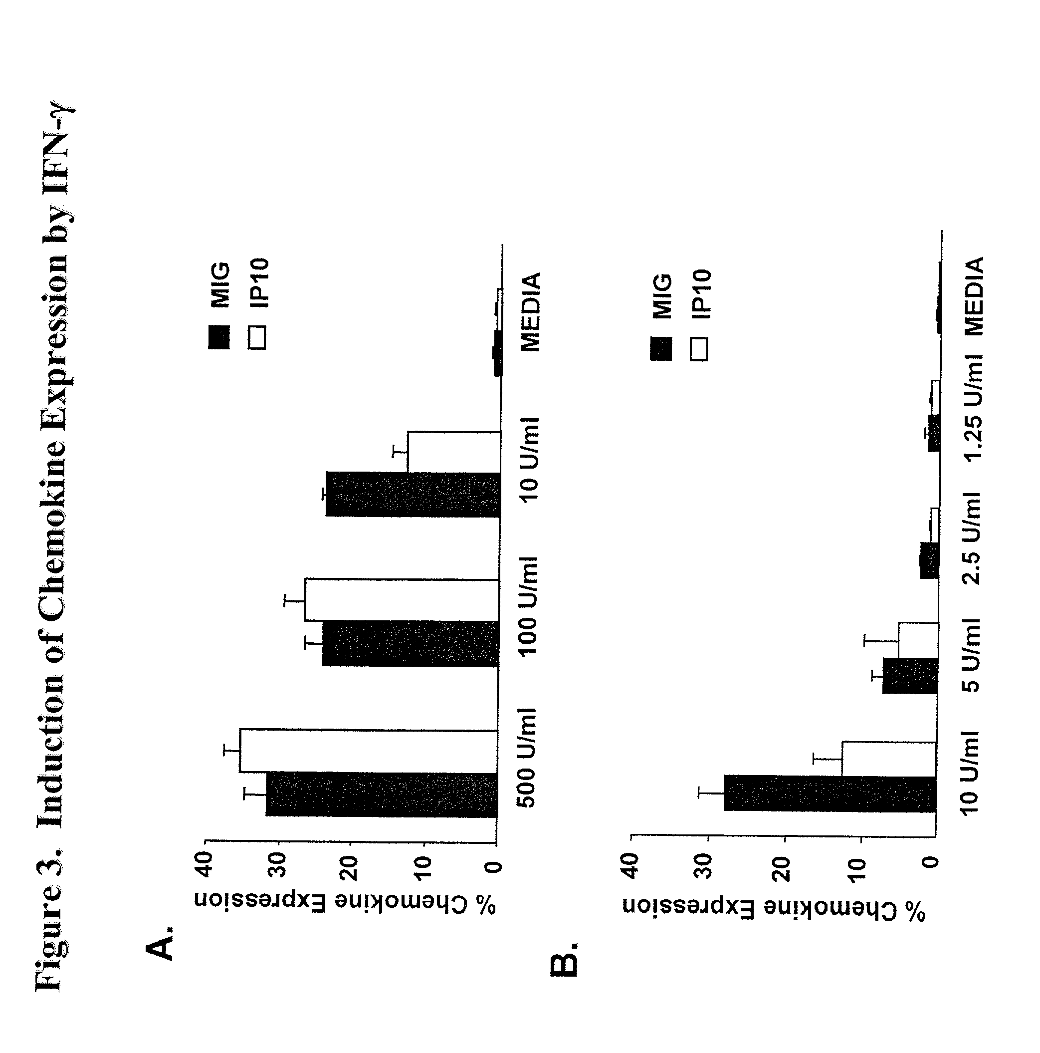
[0058] MIG is induced in response to the expression of IFN-.gamma.

[0059] To investigate the profile of chemokines induced following exposure to IFN-.gamma. which could be readily detected by intracellular staining and flow cytometry, PBMC were cultured with recombinant human IFN-.gamma. (rec.hIFN-.gamma.) and stained intracellularly with mAbs to MIG, IP-10, MCP-1, RANTES, or MIP-.alpha.. Expression of both MIG and of IP-10 was readily detectable in PBMC following overnight culture (FIG. 3A). MCP-1 and MIP-1.alpha. expression were slightly upregulated compared to media control but there was no change in expression of RANTES (data not shown). Additional dose titration experiments demonstrated that MIG expression was a more sensitive measure of IFN-.gamma. as compared to IP-10 (FIG. 3B). Moreover, it has been established that the induction of MIG is restricted to IFN-.gamma., whereas IP-10 can be induced by factors other than IFN-.gamma. including IFN-.alpha., IFN-.beta., and LPS (22-2...

example 3

[0060] Induction of MIG expression is antigen-specific and genetically restricted We evaluated whether MIG expression could be induced in an antigen-specific and genetically restricted manner. PMBC from volunteers known to express HLA-A*0201 were cultured with peptides containing HLA-A*0201-binding CD8.sup.+ T cell epitopes derived from HIV, HBV, FLU and CMV. Representative flow cytometric data from one experiment is presented in FIG. 4. MIG expression was detected with PBMC from HLA-A*0201-positive volunteers (Vols. #1,2 and 3) activated with CMV and FLU peptides, but not HIV or HBV peptides indicating that the response was antigen-specific. Additionally, antigen-specific induction of MIG expression was not detected in cultures from volunteers who did not express the HLA-A*0201 allele (Vols. #15 and #16) demonstrating that the antigen-specific response was genetically restricted. These data established that culture of PBMC with synthetic peptides elicited antigen-specific and genet...

example 4

[0061] Induction of MIG expression is IFN-.gamma. dependent

[0062] Having established that rec.hIFN-.gamma. induced MIG expression and that culture of PBMC with synthetic peptides elicited antigen-specific and genetically-restricted MIG expression, we determined whether the induction of MIG expression was dependent on IFN-.gamma.. Accordingly, PBMC were cultured with FLU or CMV peptide in the presence or absence of neutralizing mAbs to IFN-.gamma. or a control mAb (mIgG). Analysis of means and standard deviation of quadruplicate culture and staining wells is shown in FIG. 5. In two volunteers, the addition of neutralizing mAbs to IFN-.gamma. inhibited antigen-specific induction of MIG expression by an average of 87% and 85% to the FLU and CMV peptides, respectively. In repeated experiments with PBMC from volunteers 1-3, the addition of neutralizing antibodies to IFN-.gamma. inhibited antigen-specific induction of MIG expression to both FLU and CMV peptides by 86%, indicating that ant...


PUM


Abstract

Here, we describe a sensitive and specific assay and kit for the detection of chemokines having activity that is upregulated by Th-1 cytokines (such IFN-gamma) and chemokines that upregulate the activity of Th-1 cytokines (such as IFN-gamma). In a typical embodiment, detection of the chemokine monokine induced by gamma interferon (MIG) provides a measure of the biological effect of IFN-gamma rather than direct quantitation of IFN-gamma or IFN-gamma secreting cells per se. Upregulation of MIG expression was observed following in vitro activation of PBMC with defined CD8+ T cell epitopes derived from influenza virus, CMV, or EBV, and in all cases this was antigen-specific, genetically restricted and dependent on both CD8+ T cells and IFN-gamma. Responses as assessed by the MIG assay paralleled those detected by conventional IFN-gamma ELISPOT, but the magnitude of response and sensitivity of the MIG assay were superior. Our data validate this novel method for the detection of high as well as low levels of antigen-specific and genetically restricted IFN-gamma activity or MIG.

Description

[0001] 1. Field of the Invention[0002] Here, we describe a sensitive and specific assay and kit for the detection of chemokines having activity that is upregulated by Th-1 cytokines such as IFN-.gamma. (interferon-.gamma.) and chemokines that upregulate the activity of Th-1 cytokines (such as IFN-.gamma.). The invention also relates to a sensitive and novel immunoassay method and kit for detecting MIG (Monokine Induced by interferon-.gamma.) or other chemokines whose production is upregulated by IFN-.gamma.. Also, the invention relates to a method of assessing the effectiveness of a compound or system in inducing an immune response by detecting the induction of the expression of such a chemokine. All references mentioned in this application are incorporated herein by reference, in their entirety.[0003] 2. Description of the Background Art[0004] The ability to assess specific immune responses is critical for understanding the immune mechanisms underlying disease, defining the types o...

Claims


Application Information

Patent Timeline
IPC IPC(8): G01N33/50G01N33/567G01N33/68
CPCG01N33/5047G01N33/567G01N2333/57G01N33/6866G01N2333/4715G01N33/6863
Inventor BRICE, GARY TODDDOOLAN, DENISE L.HOFFMAN, STEPHEN L.
Owner THE GOVERNMENT OF THE UNITED STATES OF AMERICA AS REPRESENTED BY THE SEC OF THE NAVY NAVAL RES LAB WASHINGTON
Learn More