Nuclear transfer embryo formation method

a technology of nuclear transfer and embryo, applied in the field of nuclear transfer embryo formation method, can solve the problems of enucleation of reconstructed embryos, inability to fully enucleate cytoplasts, and inability to fully enucleate cytoplasts, and achieve the highest rate of blastocyst development, confirm comparative in vitro development potential, and increase the nt development rate

Inactive Publication Date: 2004-01-29
TRUSTEES OF TUFTS COLLEGE
View PDF5 Cites 4 Cited by
  • Summary
  • Abstract
  • Description
  • Claims
  • Application Information

AI Technical Summary

Benefits of technology

[0083] In summary, these data further confirm the comparative in vitro development potential of NT embryos produced by conventional, telophase and IE protocols. Moreover, the SIE / NT protocol appears to support the highest rate of blastocyst development when compared to the other methods. This observation is further supports the mechanism by which higher NT development rates can be obtained using the IE method. This is founded on the concept that IE affords spindle-associated enabling factors to compartmentalize within the enucleating cytoplast in a manner and timeframe so as to enhance cytoplasmic-nuclear synchronization and chromatin remodeling.

Problems solved by technology

However, physical enucleation is generally technically demanding, time consuming, inherently invasive and clearly damaging to cytoplast spatial organization.
Moreover, in certain instances, development of reconstructed embryos is inherently inefficient.
However, certain studies have reported that exposure of metaphase I and MII oocytes to etoposide, a topoisomerase II inhibitor, and cycloheximide yields enucleated cytoplasts with limited ability to support cleavage or blastocyst development, and term development of reconstructed embryos has not been reported.
However, in some instances the second PB is not fully extruded.

Method used


Image

  • Nuclear transfer embryo formation method
  • Nuclear transfer embryo formation method
  • Nuclear transfer embryo formation method

Examples


example 3

Activated Bovine Cytoplasts Produced by Induced Enucleation Support Development of Nuclear Transfer Embryos In Vitro

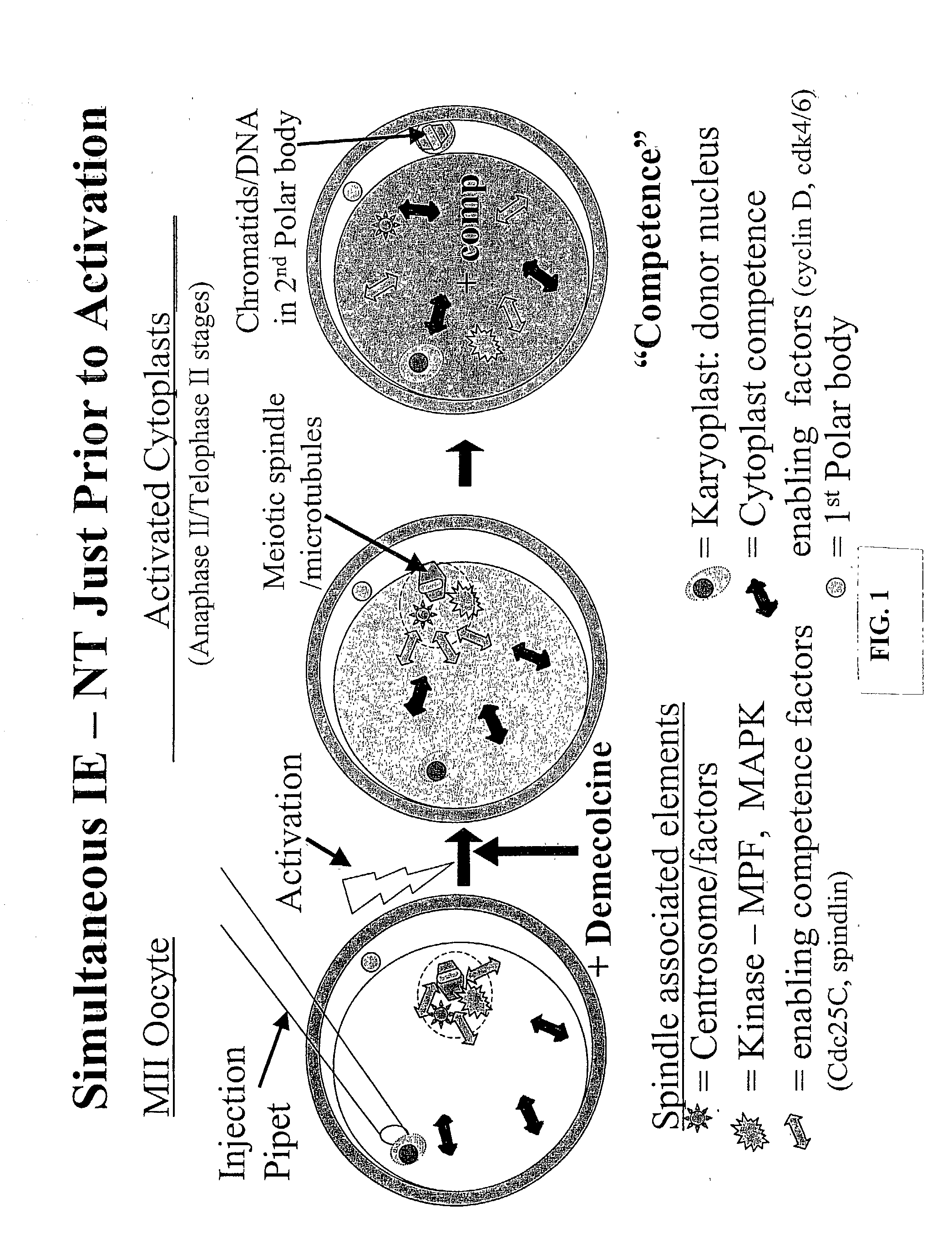
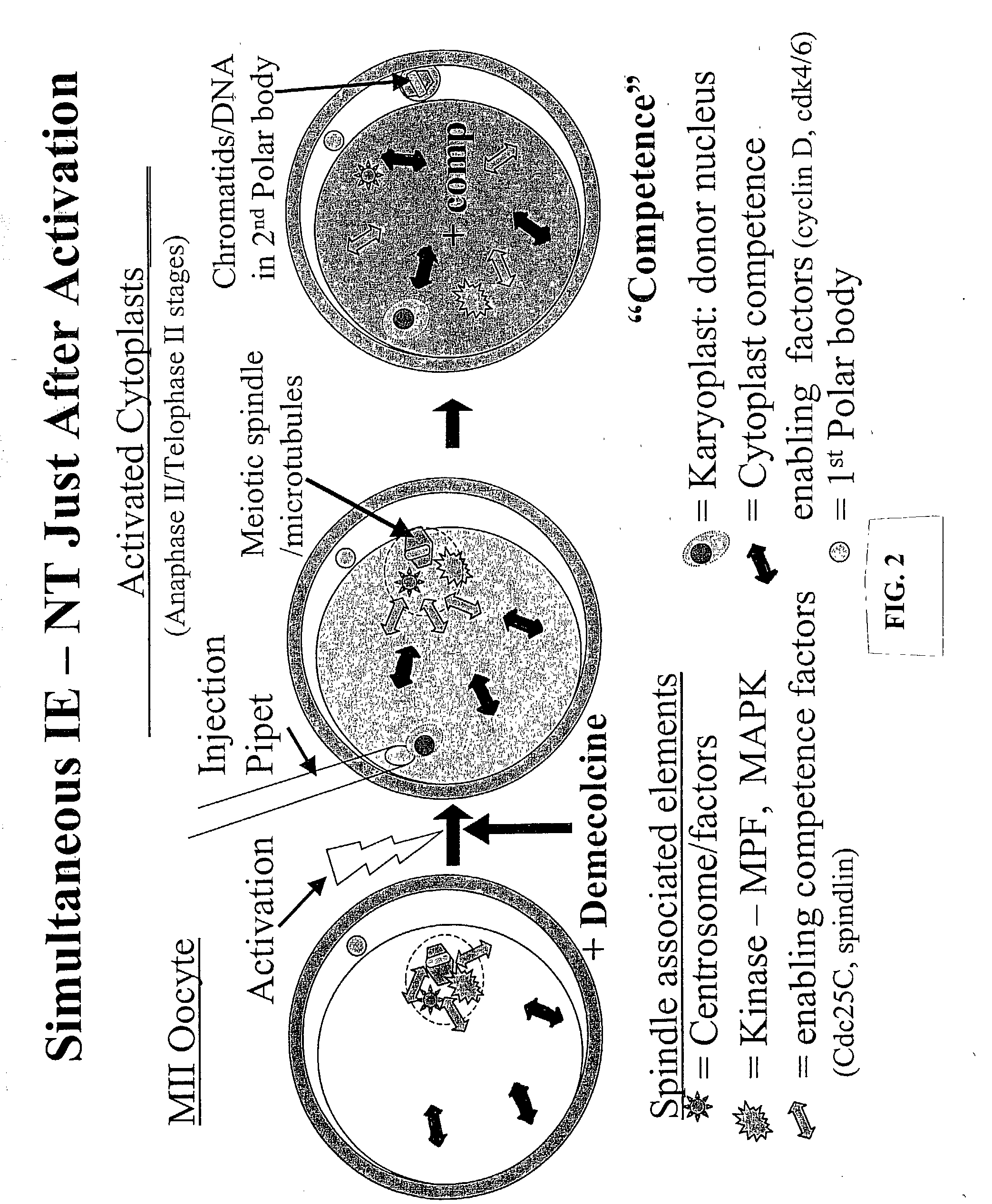
[0128] Poor efficiency of somatic cell NT has been associated with the preparation of developmentally competent enucleated cytoplasts. Induced enucleation (IE) of mouse oocytes has been shown to support enhanced term development of cloned mice. This study characterized the kinetics and phenotypic progression of bovine oocytes subjected to IE, and evaluated their developmental competence to support NT embryo development in vitro. In vitro matured (26 h) oocytes were denuded, activated (5 pM ionomycin, 5 min, then 10 .mu.g / mL cycloheximide, 5 h) and cultured for up to 5 h post-activation (pa). Oocyte enucleation was induced by demecolcine (0.4 .mu.g / ml, DM) exposure at 30, 60, 90 and 120 min post activation for various time periods (1 to 4.5 h). Activation rates and meiotic progression of control and DM treated oocytes (n=31-49 / gp) was evaluated at 5 hpa by immunofluores...

example 4

Induced Enucleation of Mouse and Goat Oocytes Kinetic and Phenotypic Characterizations

[0130] In general, the rates of successful somatic cell cloning of animals are poor, in part due to low efficiency in the production of competent cytoplasts when prepared by mechanical enucleation of MII oocytes. A method to induce enucleation of activated oocytes has been developed that supports enhanced rates of development of cloned mice to term. This study was designed to characterize the kinetics and progressive phenotypes observed during induced enucleation (IE) of activated mouse and goat oocytes. In vivo ovulated mouse oocytes (B6D2F1, n-959; CF 1, n=999) and in vitro matured goat oocytes (n-163) were denuded activated (7% ethanol) and incubated for up to 3.5 hr in KSOM. Enucleation was induced by continuous exposure to demecolcine (0.4 g / ml) commencing at 0-30 min post-activation. Non-demecolcine treated activated oocytes served as controls (n=50 / rep). At selected time points oocytes (n=11...


PUM

PropertyMeasurementUnit
time-aaaaaaaaaa
time-aaaaaaaaaa
time-aaaaaaaaaa

Abstract

A nuclear transfer embryo is formed by destabilizing microtubules of an oocyte, whereby essentially all endogenous chromatin collects at a second polar body during meiosis of an oocyte. The oocyte is fused with the nucleus of a donor somatic cell of the same species of said oocyte prior to cessation of extrusion of the second polar body from the oocyte, thereby forming the nuclear transfer embryo. In one embodiment, the nuclear transfer embryo is employed to impregnate an animal, such as a mammal. In another embodiment, the donor nucleus is transgenic.

Description

[0002] Nuclear transfer methods have been developed and used successfully to produce cloned sheep, cattle, mice, goats and pigs. Two cell components are combined to produce a cloned embryo; the donor nuclear genome (karyoplast) that is the target for clonal replication, and the enucleated oocyte (cytoplast) whose cytoplasmic constituency is sufficiently competent to facilitate genome reprogramming and support embryonic development to term.[0003] Mammalian oocyte cytoplasts have been prepared by physically removing nuclear chromatin by micromanipulation techniques in preparation to receive the donor genome. Enucleated oocytes arrested at metaphase of meiosis II (MII) are subsequently "reconstructed" by the addition of the donor karyoplast typically using either electrofusion or microinjection techniques. However, physical enucleation is generally technically demanding, time consuming, inherently invasive and clearly damaging to cytoplast spatial organization. Moreover, in certain ins...

Claims


Application Information

Patent Timeline
Patent Type & Authority Applications(United States)
IPC IPC(8): C12N15/873C12N15/877
CPCA01K2227/101C12N15/8771C12N15/873
Inventor OVERSTROM, ERIC W.FISCHER, DANIELA
Owner TRUSTEES OF TUFTS COLLEGE
Why Eureka
Try Eureka