
Detection of chemical ligation using fluorescence quenching leaving groups

a technology of quenching and chemical ligation, which is applied in the field of detection of chemical ligation, can solve the problems of false positives, dangerously long bacterial culture period, and non-specific binding of target nucleic acids to oligonucleotide probes to target dna,

Inactive Publication Date: 2004-12-23
SANDFORD UNIV +1
View PDF10 Cites 30 Cited by
  • Summary
  • Abstract
  • Description
  • Claims
  • Application Information

AI Technical Summary

Benefits of technology

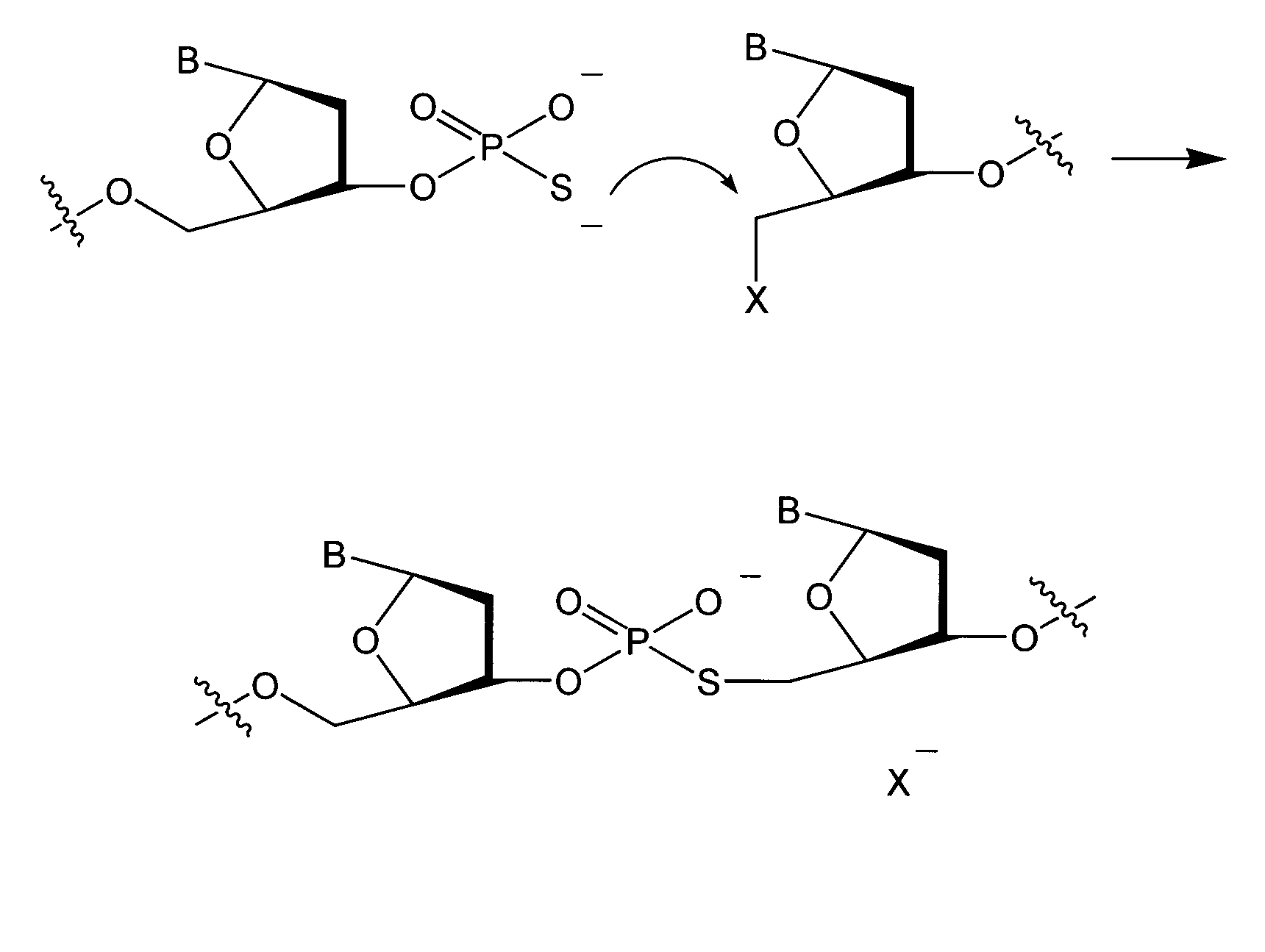
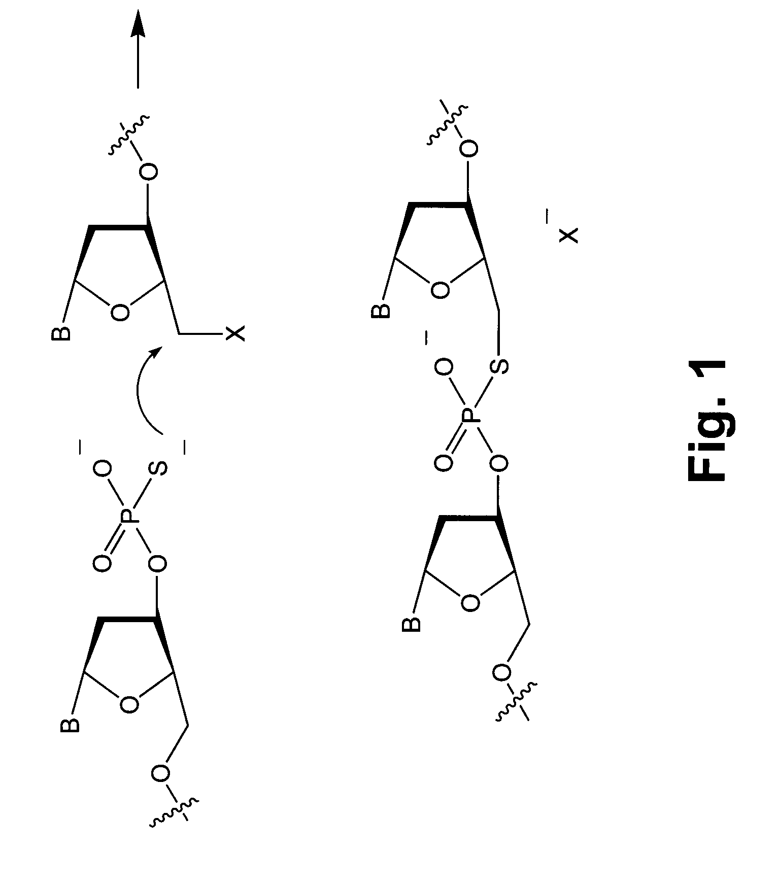
0021] Oligonucleotides containing a fluorophore and a specialized quencher that also acts as a leaving group are disclosed. Nucleophilic attack on the quenched DNA, causing release of the quencher group, results in a liga

Problems solved by technology

Even short (e.g. two days) bacterial cultures are dangerously long for patients with other acute infections such as those occurring in sepsis or in necrotizing fasciitis.
One limitation in using the standard hybridization method for detecting target nucleic acids is non-specific binding of the oligonucleotide probes to the target DNA or RNA.
Despite this improvement to the standard hybridization method, false positives may still result from the oligonucleotide probes non-selectively binding to proteins or the solid support.
Standard hybridization methods using static labeling groups are further limited in that they usually have to be performed on solid supports under stringent conditions and require careful washing (static labeling here refers to labels, such as fluorescent labeles, that do not change their signal).
Thus, standard hybridization methods using statically labeled oligonucleotides require time for preparation of the cells, increase the likelihood of error, and cannot be used in live cells, where washing away unbound probes is not possible.
However, this method is limited because the fluorescence change clearly depends on solution conditions, e.g. temperature, ionic strength, and thus conditions must be monitored carefully.
Another disadvantage is that molecular beacon method is not as sequence selective as other DNA-sensing methods such as enzymatic approaches or some non-enzymatic autoligation methods.
When the molecular beacon approach was recently used to image RNA in live mammalian cells, the results were disputed because these probes can give false positives by being degraded or by binding a protein instead of RNA.
There are many DNA-and RNA-binding proteins in a cell, so false positives are likely due to nonspecific binding of the probes.
However, the reported specificity is not as high as phosphorothioate-iodide autoligation probes.
As described above, such beacons suffer from false positives by binding proteins.

Method used


Image

  • Detection of chemical ligation using fluorescence quenching leaving groups
  • Detection of chemical ligation using fluorescence quenching leaving groups
  • Detection of chemical ligation using fluorescence quenching leaving groups

Examples


example 1

[0054] Reaction in Solution

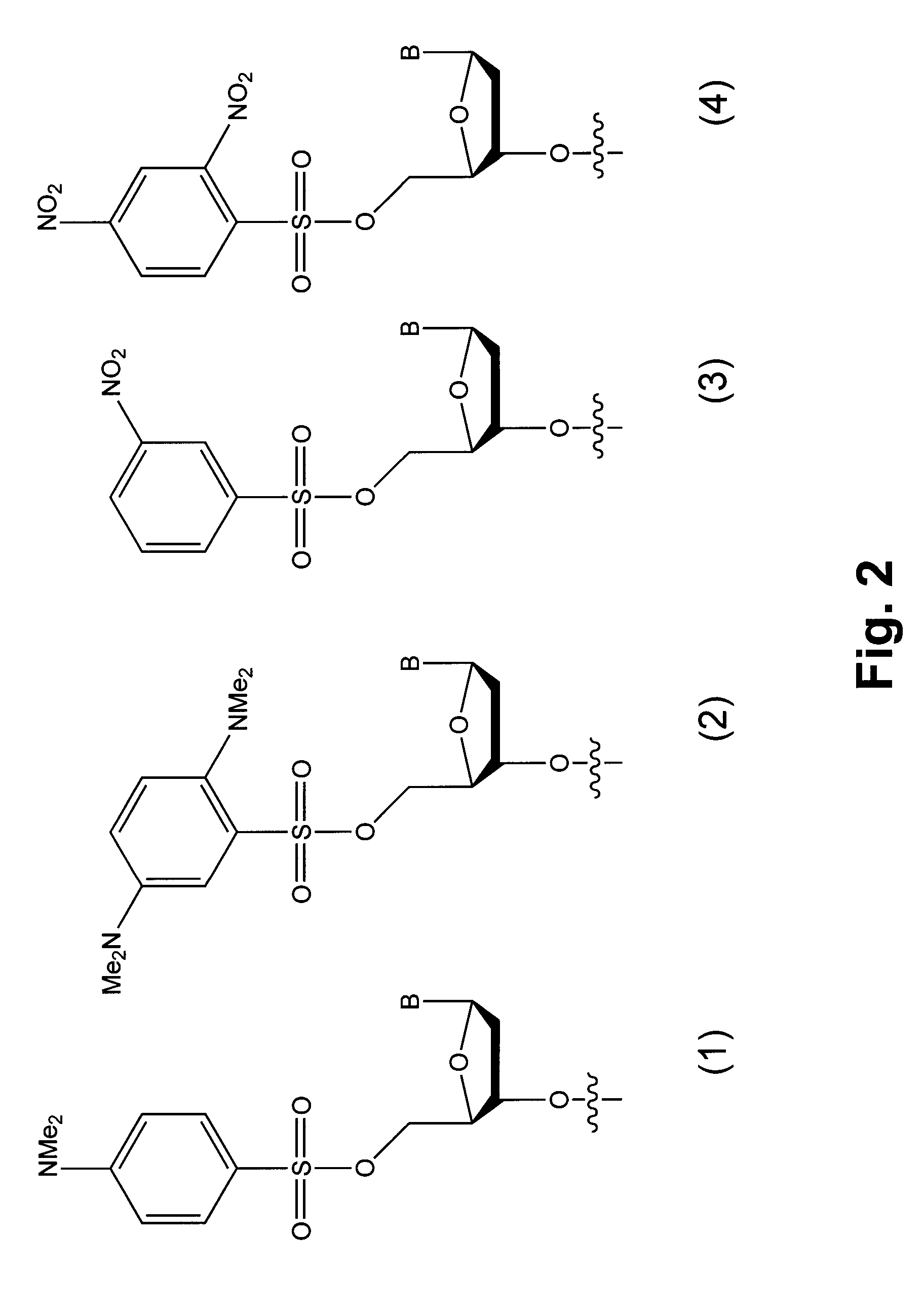
[0055] Wild type and mutant sequences 50 nucleotides in length were chosen from the H-ras gene. A known C to A transversion in codon 12 was selected to fall at the center of a 7mer binding site. The nucleophilic 7mer sequence was 5'-CCGTCGG-3' (SEQ ID NO:4), where the central T hybridizes to the A transversion, but not to the wild type C. The nucleophilic sequence contained a 3' phosphorothioate group. The electrophilic 13mer sequence was 5'-TGT*GGGCAAGAG-3' (SEQ ID NO: 1). A dabsyl group was used on the 5' hydroxyl as a leaving group of the electrophilic sequence, and a commercially available fluorescein C-5-alkenyl conjugate of dU (marked as T*) was used to place the fluorophore two nucleotides away from the quencher. The wild type target sequence was 3'-ATATTCGACCACCACCACCCGCGGCC-GCCACACCCGTTC TCACGCGACTG-5' (SEQ ID NO:2), and the mutant transversion target sequence was 3'-ATATTCGACCACCACCACCCGCGGCAGCCACACCCGTTC TCACGCGACTG-5' (SEQ ID NO:3; where the C ...

example 2

[0058] Reaction on a Solid Support

[0059] As many genetic analysis methods use probes affixed to beads, slides, and other surfaces, a solid support was used to evaluate the instant invention. A 7mer MUT probe (5'-CCGTCGG-3'; SEQ ID NO:4) on 90 .mu.m beads (1000A pore size) using commercially available reverse (5'.fwdarw.3') phosphoramidites was used, placing a 3' phosphorothioate moiety on the final 3' hydroxyl group. A hexaethylene glycol linker was used to alleviate potential crowding problems near the polymer surface. Such beads then have the potential to autoligate a 13mer quenched electrophile probe to themselves, in the presence of the correct target DNA. This was expected to result in the beads becoming fluorescent, as the dabsylate group was lost and the nearby fluorescein label lost quenching. The solid-phase autoligations were monitored by imaging under a fluorescence microscope. Results showed that the reaction proceeded on the polystyrene beads much as it does in solution...

example 3

[0060] Use of TAMRA as a Fluorophore

[0061] Example 1 was repeated using a different fluorophore. The use of different fluorophores would make it possible to sense multiple genetic sequences simultaneously. Dabsyl has been reported as a quencher for varied fluorophores. The same ligations on beads were carried out using a dabsyl / TAMRA electrophilic probe. The results showed that ligation and unquenching was also successful for the new dye.


PUM

PropertyMeasurementUnit
Efficiencyaaaaaaaaaa
Fluorescenceaaaaaaaaaa

Abstract

Novel compounds having a fluorescence quencher as a leaving group are disclosed. Nucleic acids and other molecules containing a fluorophore and a fluorescence quencher are disclosed as an embodiment of this invention. The use of the oligonucleotides in enzyme-free oligonucleotide ligation reactions results in an increase in fluorescence when the oligonucleotide also contains a nearby fluorophore. The ligation reactions can be performed in solution, on surfaces, or in cells.

Description

[0001] The present application claims priority to U.S. Provisional Patent Application Ser. No. 60 / 396,774 filed Jul. 18, 2002, the contents of which are incorporated herein by reference.BACKGROUND OF INVENTION[0003] 1. Field of the Invention[0004] The invention relates to methods of detecting chemical ligation. One example application involves methods of detecting nucleic acids and, more specifically, to methods for the detection of ligation of nucleic acids via a change in fluorescence properties. Methods for the detection of ligation by the use of a quencher as a leaving group are disclosed.[0005] 2. Description of the Related Art[0006] As the molecular nature of diseases is studied, there is an increasing need for methods of detecting and analyzing nucleic acids both in vitro and in vivo. Recent work on sequencing the human genome has resulted in a flood of genomic and proteomic information. This information has made a significant and sometimes life-determining difference in the ...

Claims


Application Information

Patent Timeline
IPC IPC(8): C07H21/04C07K14/00C12Q1/68G01N33/53G01N33/533G01N33/542G01N33/58
CPCC12Q1/6818C12Q1/6823C12Q1/6827G01N33/533G01N33/542C12Q2561/125C12Q2565/101
Inventor KOOL, ERIC T.
Owner SANDFORD UNIV
Learn More