Minimally invasive diagnosis and treatment for breast cancer

a breast cancer, minimally invasive technology, applied in the field of cancer treatment, can solve the problems of affecting the detection affecting the accuracy of the lesion recognition, so as to achieve the effect of reducing the fluorescen

Inactive Publication Date: 2005-01-06
INNOBLATIVE DESIGNS INC
View PDF24 Cites 25 Cited by
  • Summary
  • Abstract
  • Description
  • Claims
  • Application Information

AI Technical Summary

Benefits of technology

[0032] The present invention is directed further to a method of treating a close or positive margin of an excisional site of a cancerous lesion in a breast of an individual. The close or positive margin of the excisional site is located via imaging thereof whereupon a fluorophore is administered intravascularly to the individual and the fluorescence of the fluorophore is measured at the excisional site. A second margin is ablated with a radiofrequency probe under guided imaging and fluorescence of the fluorophore is measured as the second margin is ablated. A decrease in the fluorescence correlates with an increase in ablation of the close or positive margin to form a second cancer free margin thereby treating the close or positive margin.

Problems solved by technology

Visibility of the lesion by the imaging system may be hampered because of the distortion created by the extraction process itself as well as associated bleeding in the surrounding tissues.
Although the lesion is removed and all fluids are continuously aspirated from the extraction site, it is likely that the process will “cloud” the lesion, thus impairing exact recognition of its margins.
This makes it difficult to ensure that the entire lesion will be removed.
Such a lesion actually serves to mark or define the location of adjacent abnormal tissue, so the physician does not wish to remove the entire lesion and, thereby, lose a critical means for later relocating the affected tissue.
Thus, if subsequent analysis indicates the tissue to be malignant, it is difficult for the physician to determine the precise location of the lesion in order to perform necessary additional procedures on adjacent potentially cancerous tissue.
While such a location system is effective, it is obviously intended and designed to be only temporary and is removed once the surgery or other procedure has been completed.
Obviously, however, none of these approaches are useful for marking and delineating internal tissue abnormalities, such as lesions, tumors or margins.
Of course, this method also is only a temporary means for marking the abnormal tissue.
Additionally, once the abnormal tissue has been destroyed during treatment, the marker is destroyed as well.
However, while such staining techniques can be effective, it is difficult to precisely localize the stain.
Also, the stains are difficult to detect using fluorescence imaging and may not be permanent.
Thermally-induced apoptosis impairs adequate pathologic assessment of tumor grade, lymphovascular invasion and biologic tumor markers, i.e., estrogen, progesterone and HER-2 / neu, therefore necessitating a pre-ablative size assessment and histologic diagnosis via core bipsy.
The limitation on lesion size is due to the range of zone of ablation created by the laser.
This agitation results in frictional heat to ultimately induce coagulative necrosis.
Incomplete ablation in four patients was thought due to the inability to adequately size the tumor with standard preoperative imaging or secondary to inappropriate targeting via ultrasound.
However, prior to surgery, the patients must wait for pathology results of a biopsy before a final diagnosis can be given.
Moreover, breast conservation surgery via lumpectomy often results in some deformity and requires repeated surgery to establish a clear margin around the malignant lesion.
None of the presently available devices for excision adequately excise a margin, i.e., a tumor-free zone.
Additionally, no work has been published on ablation of tumor bed margin after excision.
Specifically, the prior art is deficient in methods of treating breast cancer by excising the tumor and ablating the margin of the tumor in a minimally invasive manner.

Method used


Image

  • Minimally invasive diagnosis and treatment for breast cancer
  • Minimally invasive diagnosis and treatment for breast cancer
  • Minimally invasive diagnosis and treatment for breast cancer

Examples


example 1

[0068] MRI-directed Interstitial Laser Photocoagulation

[0069] Preliminary results of MRI-directed interstitial laser photocoagulation in 30 patients with breast cancer indicated that the procedure was well-tolerated and side effects were similar to those of routine stereotactic needle biopsy. Each patient has between one and five ablation zones for a total of 68 treatment zones. When the entire 10-minute treatment session was completed, the pathology correlation determined effective cell death in all cases. The histologic determination of treatment zones by proliferating cell nuclear antigen (PCNA) staining within the tumor matched the MRI estimates of zone size in all cases (data not shown).

example 2

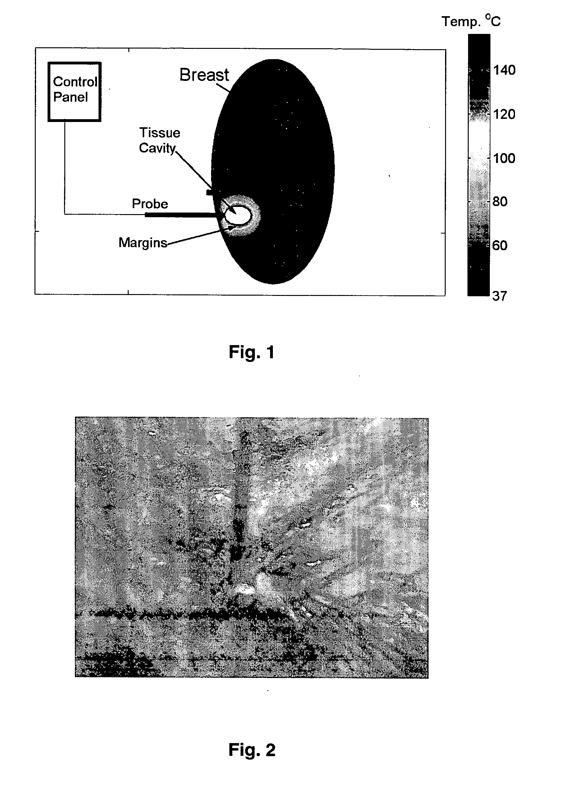
[0070] Excision and Ablation of Margin in Mastectomy Specimens

[0071] Normal tissue was excised, under IRP-approved protocol, from 39 sites on mastectomy specimens. The sites were then ablated with radiofrequency to measure the margin produced around the cavity site (FIG. 2).

[0072] Tumors were excised percutaneously from 10 patients using the Mammotome®. The margin was ablated and then the site was excised for pathological examination. Adequate margins were obtained on all specimens examined (data not shown).

example 3

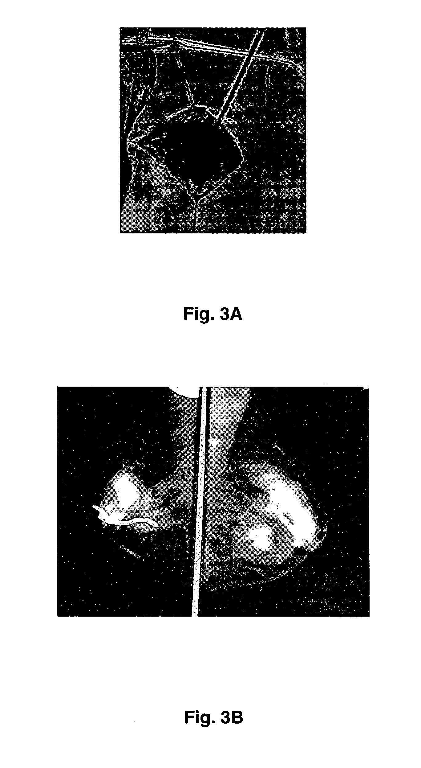
[0073] Excision and Ablation of Margin as Alternative to Excision and Radiation Therapy

[0074] Because of debilitation of the patient and / or refusal of the patient to undergo radiation therapy, the treatment protocol of excision and radiation therapy is not a viable procedure. In 10 such patients, standard excision followed by ablation of the margin with radiofrequency was performed (FIG. 3A). The patients were followed-up closely over a 2-year period or less with no recurrences in any of them.

[0075] In one such instance an elderly woman with severe pulmonary disease who had a large right breast cancer removed was treated with radiofrequency ablation and follow-up. This patient demonstrates none of the typical visible side-effects of radiation therapy including retraction, swelling, erythema or browning of the skin. Two year mammograms show no evidence of recurrence (FIG. 3B). Two-year follow-up is emphasized since most recurrences for breast tumors occur in the first 2 years after...


PUM


Abstract

The present invention provides a treatment method to excise a cancerous lesion, such as in the breast, with subsequent ablation of the margin. The method provides for location and excision with ablation under open guidance or guided imaging and for diagnosis by cytology. The method may be a minimally, invasive same day method with diagnosis before or immediately after excision. Also provided is a method of treating close or positive margins of an excisional site of a cancerous lesion in a breast by ablating the margin while monitoring the fluorescence of a fluorophor at the site to determine when ablation of the close or positive margin has occurred.

Description

CROSS-REFERENCE TO RELATED APPLICATION [0001] This application is a continuation-in-part of non-provisional U.S. Ser. No. 10 / 306,117, filed Nov. 27, 2002, which claims benefit of provisional U.S. Ser. No. 60 / 337,626, filed Dec. 10, 2001, now abandoned.BACKGROUND OF THE INVENTION [0002] 1. Field of the Invention [0003] The present invention relates generally to the field of cancer treatment. More specifically, the present invention relates to a method of diagnosis and treatment for cancer such as breast cancer by combining techniques of tumor excision and margin ablation. [0004] 2. Description of the Related Art [0005] Breast cancer will be diagnosed in approximately one in eight women in their lifetime and one in 30 will die of this disease. It is the leading cause of cancer deaths in women 40-55 years of age and the second leading cause of cancer deaths in women overall. Breast cancer does occur in males, but is much less common. [0006] It is desirable and often necessary to perfor...

Claims


Application Information

Patent Timeline
Patent Type & Authority Applications(United States)
IPC IPC(8): A61B10/00A61B10/02A61B17/00A61B18/24A61B19/00
CPCA61B10/0041A61B10/0233A61B2019/5236A61B2017/008A61B2018/2005A61B10/0283A61B2090/374
Inventor KLIMBERG, V. SUZANNEKOROURIAN, SOHEILAHARMS, STEVEN
Owner INNOBLATIVE DESIGNS INC
Why Eureka
Try Eureka