Method and system for ultrasonic tagging of fluorescence

a fluorescence and ultrasonic technology, applied in the field of medical imaging systems, can solve the problems of later stage detection of disease, low sensitivity of the procedure, and inability to precisely detect radiologically dense breasts, so as to reduce the computational time for reconstruction and improve spatial resolution

Inactive Publication Date: 2005-05-19
GENERAL ELECTRIC CO
View PDF10 Cites 116 Cited by
  • Summary
  • Abstract
  • Description
  • Claims
  • Application Information

AI Technical Summary

Benefits of technology

[0009] A method and system for localization of fluorescent dyes in a scattering medium such as a biological tissue are provided. In comparison to other optical imaging techniques, this d

Problems solved by technology

The specificity of this exam for radiologically dense breasts is not optimal.
The low sensitivity of the procedure gives rise to a high number of false negative results.
This, in turn, leads to later stage detection of the disease and poorer patient prognostics.
However, the increased frequency of mammography procedures, combined with the risks associated with the increased exposure to ionizing radiation brings into focus the clinical need for an alternate screening procedure that

Method used


Image

  • Method and system for ultrasonic tagging of fluorescence
  • Method and system for ultrasonic tagging of fluorescence
  • Method and system for ultrasonic tagging of fluorescence

Examples


Embodiment Construction

[0022] Preferred embodiments of the present invention will be described hereinbelow with reference to the accompanying drawings. In the following description, well-known functions or constructions are not described in detail to avoid obscuring the invention in unnecessary detail.

[0023] A system and method for the localization of fluorescent dyes in a scattering medium are provided. In various embodiments of the present invention, systems and methods will be employed for localizing an object of interest, e.g., a lesion labeled with a fluorescent dye, in a scattering medium, e.g., biological tissue.

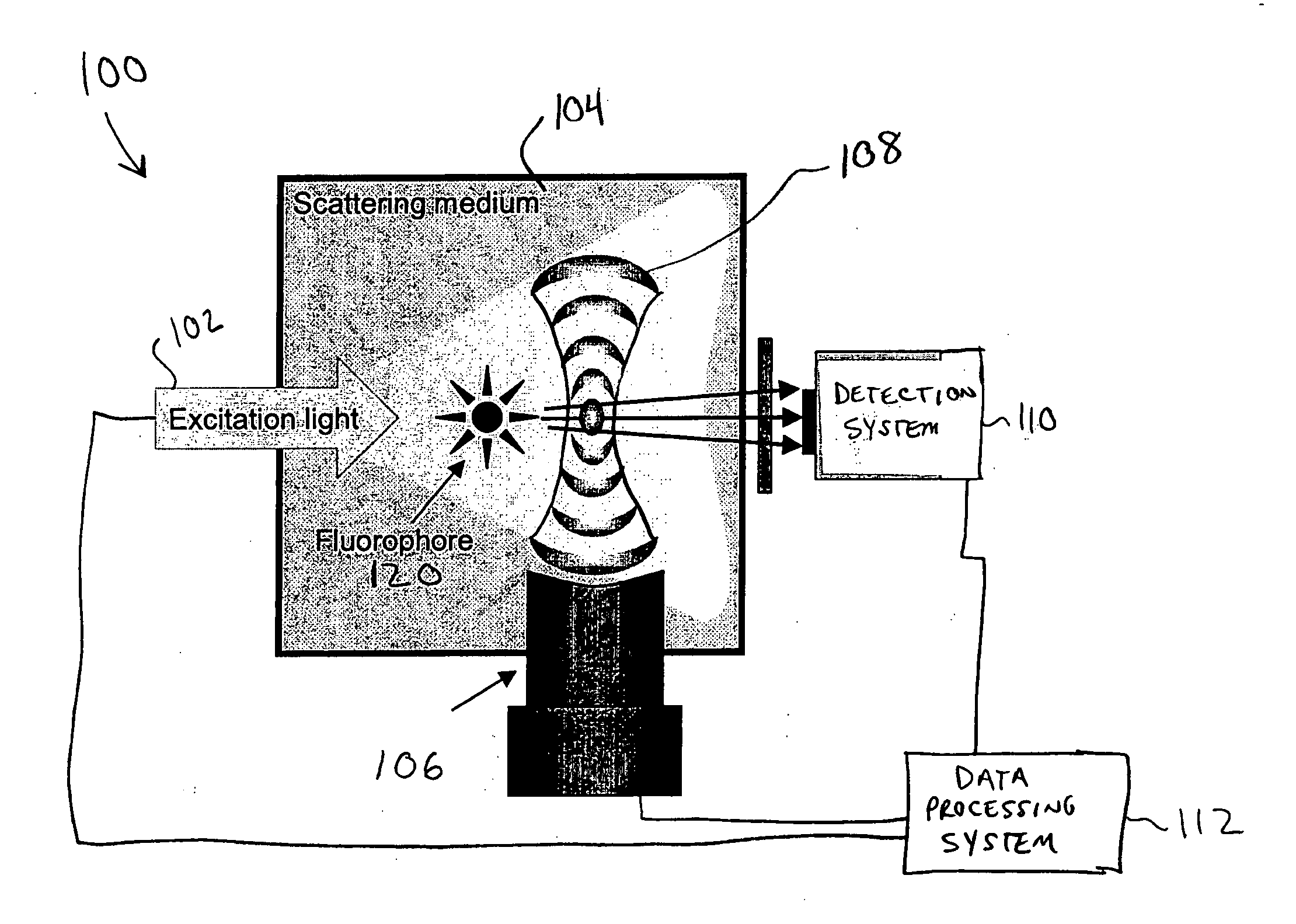
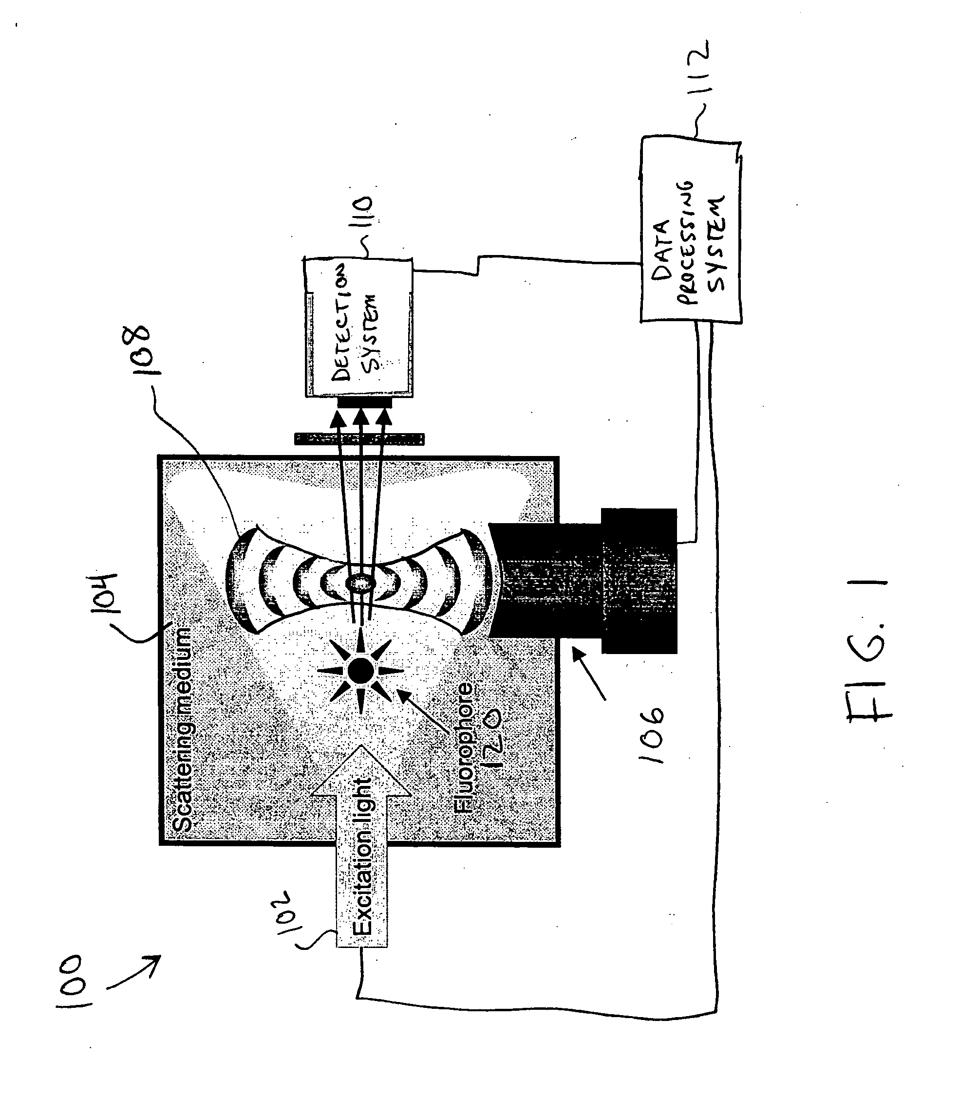
[0024] Referring to FIG. 1, the system 100 generally includes an optical excitation source 102 for irradiating or illuminating a scattering medium 104, including an object of interest 120, with radiant energy, e.g., light. The light excites a fluorophore contrast agent present in the object of interest 120. The system 100 further includes an ultrasonic generation system 106 for generating...


PUM


Abstract

A method and system for localization of fluorescence in a scattering medium such as a biological tissue are provided. In comparison to other optical imaging techniques, this disclosure provides for improved spatial resolution, decreased computational time for reconstructions, and allows anatomical and functional imaging simultaneously. The method including the steps of illuminating the scattering medium with an excitation light to excite the fluorescence; modulating a portion of the emitted light from the fluorescence within the scattering medium using an ultrasonically induced variation of material properties of the scattering medium such as the refractive index; detecting the modulated optical signal at a surface of the scattering medium; and reconstructing a spatial distribution of the fluorescence in the scattering medium from the detected signal.

Description

BACKGROUND OF THE INVENTION [0001] This disclosure relates generally to medical imaging systems, and more particularly, to methods and systems for localization of fluorescent dyes in a scattering medium, e.g., biological tissue. [0002] Millions of women participate in x-ray mammography each year. The specificity of this exam for radiologically dense breasts is not optimal. Suspicious lesions are subject to follow-up with other modalities such as ultrasound imaging, and ultimately are likely to be examined by biopsy. The physical discomfort and psychological impact to the women who obtain false-positive results from this screening procedure is immeasurable. The low sensitivity of the procedure gives rise to a high number of false negative results. This, in turn, leads to later stage detection of the disease and poorer patient prognostics. The rate of women having x-ray mammography screening is increasing annually, which leads to an overall earlier detection of suspect lesions. Howeve...

Claims


Application Information

Patent Timeline
IPC IPC(8): A61B5/00G01N21/17G01N21/47
CPCA61B5/0048A61B5/0091A61B5/4312A61K49/0017A61K49/0032A61K49/0034G01N2021/6439B82Y10/00B82Y20/00G01N21/1717G01N21/4795G01N21/6456G01N2021/1729B82Y5/00A61B5/0051
Inventor JANSEN, FLORIBERTUS HEUKENSFELDTLORRAINE, PETER WILLIAMLOMNES, STEPHEN JOHNSONFOMITCHOV, PAVEL ALEXEYEVICH
Owner GENERAL ELECTRIC CO
Why Eureka
Try Eureka