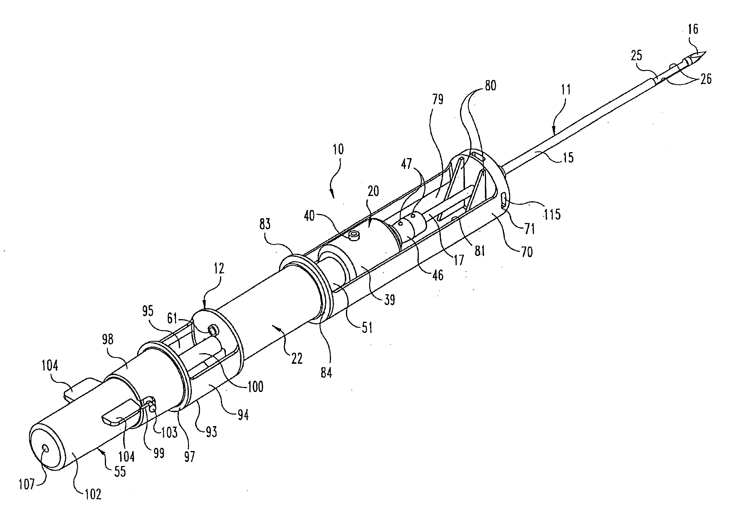
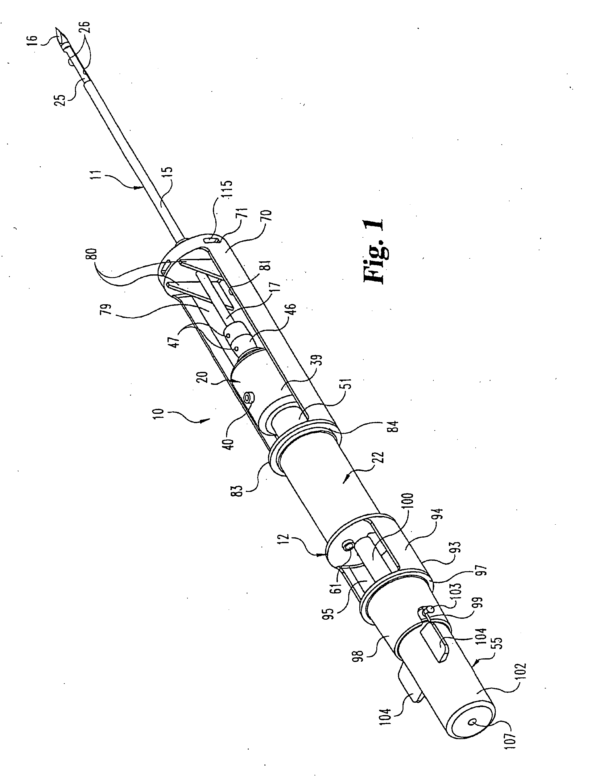
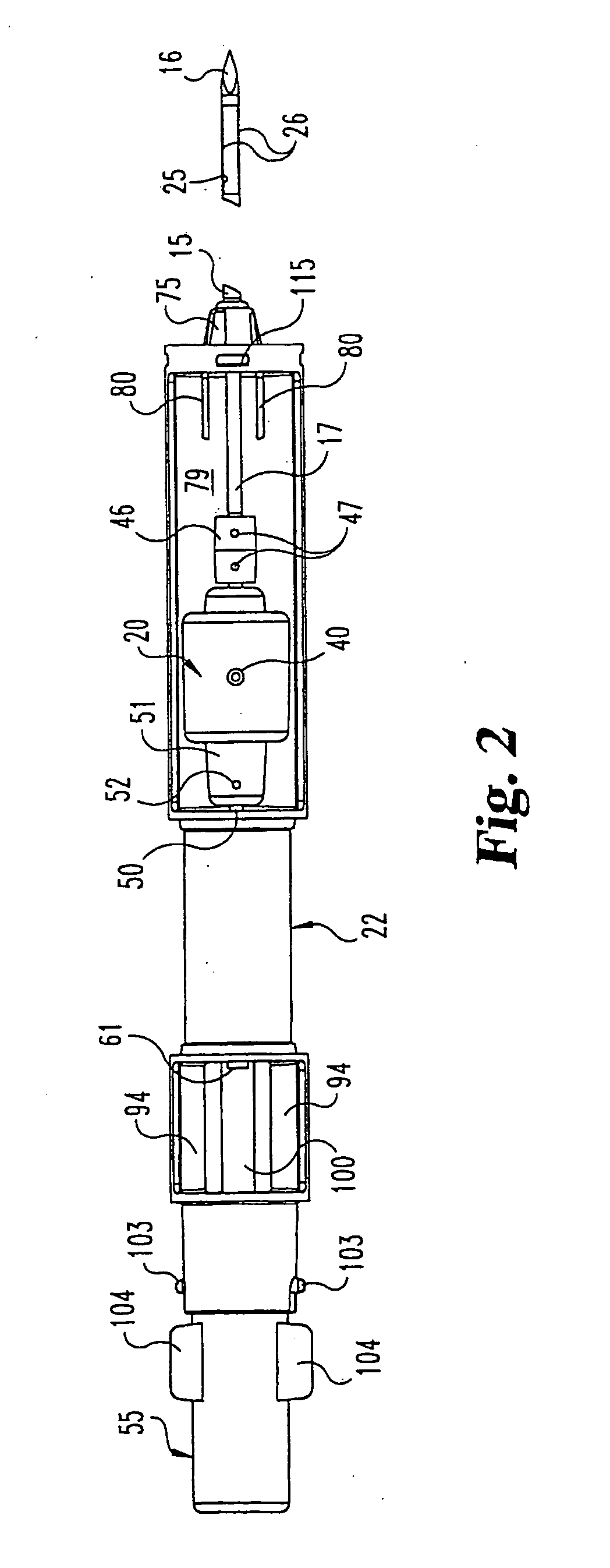
Biopsy apparatus

a biopsy apparatus and apparatus technology, applied in the field of biopsy instruments, can solve the problems of increasing the risk of infection and bleeding at the sample site, significant trauma to the breast tissue, and requiring considerable recovery time for the patient, so as to achieve the effect of maximizing the length and overall size of the core, avoiding infection and bleeding, and ensuring the integrity of longitudinal integrity

Inactive Publication Date: 2005-05-26
TISSUE EXTRACTION DEVICES
View PDF89 Cites 292 Cited by
  • Summary
  • Abstract
  • Description
  • Claims
  • Application Information

AI Technical Summary

Benefits of technology

[0023] The outer cannula has a trocar tip at its distal end and a cutting board snugly disposed within the outer cannula. The inner cannula defines an inner lumen that extends the length of the inner cannula, and which provides an avenue for aspiration. The inner cannula terminates in an inwardly beveled, razor-sharp cutting edge and is driven by, both a rotary motor, and a reciprocating motor. As the inner cannula moves past the tissue-receiving opening, the inwardly beveled edge helps to eliminate the risk of catching the edge on the tissue-receiving opening. At the end of its stroke, the inner cannula makes contact with the cutting board to completely sever the tissue. The cutting board is made of a material that is mechanically softer than the cutting edge yet hard enough to withstand the force of the inner cannula.
[0026] In another embodiment, the tissue-receiving opening is formed by opposite longitudinal edges that form a number of teeth. The teeth face away from the cutting board at the distal end of the outer cannula. The teeth help prevent the forward motion of the tissue in the opening as the inner cannula moves forward toward the cutting board. This feature maximizes the length and overall size of the core, ultimately resulting in a more efficient lesion removal.
[0027] In another embodiment, the outer cannula incorporates a stiffening element opposite the tissue-receiving opening. This stiffening element aids in maintaining the longitudinal integrity of the outer cannula as it is advanced through the tissue.
[0028] In addition to the inwardly beveled edge of the inner cannula, one embodiment incorporates additional features to prevent the inner cannula from rising up into the tissue-receiving opening. A bead of stiffening material may be affixed to the inner wall of the outer cannula, or a dimple may be formed in the inner wall of the outer cannula. The bead, or dimple urges the inner cannula away from the tissue-receiving opening and prevents the inner cannula from catching on the opening.

Problems solved by technology

This procedure causes significant trauma to the breast tissue, often leaving disfiguring results and requiring considerable recovery time for the patient.
This is often a deterrent to patients receiving the medical care they require.
The open technique, as compared to the percutaneous method, presents increased risk of infection and bleeding at the sample site.
In addition, this method does not provide for a pathological assessment of the tissue, which can provide a more complete assessment of the stage of the cancer, if found.
First, the device would sometimes fail to remove a sample, therefore, requiring additional insertions.
This was generally due to tissue failing to prolapse into the sampling notch.
Secondly, the device had to be inserted and withdrawn to obtain each sample, therefore, requiring several insertions in order to acquire sufficient tissue for pathology.
Although the Burbank device presents an advancement in the field of biopsy devices, several disadvantages remain and further improvements are needed.
These small holes often become clogged with blood and bodily fluids.
This increases the time necessary to wrap up the procedure, which ultimately affects the cost of the procedure.
In addition, the required clean-up and / or sterilization of reusable parts increases the staffs' potential exposure to body tissues and fluids.
Finally, the reusable handle is heavy, large and cumbersome for handheld use.
A further disadvantage is that current biopsy devices comprise an open system where the tissue discharge port is simply an open area of the device.
In addition, the open system increases the exposure to potentially infectious materials, and requires increased handling of the sample.
As a practical matter, the open system also substantially increases the clean-up time and exposure, because a significant amount of blood and bodily fluid leaks from the device onto the floor and underlying equipment.
Additionally, when using the current biopsy devices, physicians have encountered significant difficulties severing the tissue.
For instance, the inner cutter often fails to completely sever the tissue.
In the case of the Burbank apparatus, the failure to completely sever the tissue after the first advancement of the inner cutter results in a necessary second advancement of the inner cutter.
In this event, the procedure is prolonged, which is significant because the amount of trauma to the tissue and, ultimately, to the patient is greatly affected by the length of the procedure.
This lifting causes the inner cutter to catch on the edge of the tissue receiving opening, which ultimately results in an incomplete cut and dulling of the blade, rendering the blade useless.
Also, prior devices often produce small tissue samples.
An additional disadvantage of the prior devices is presented by the complexity of the three-pedal footswitch.
Operation of the three pedals is difficult and awkward.
These disadvantages become even more significant when using the handheld biopsy device.
For instance, the physician must operate the biopsy device and the ultrasound probe simultaneously making it particularly difficult to manually advance of the inner cutter.
In addition, when an assistant is required to remove each sample from the open discharge port, use of the handheld device becomes even more awkward.
Due to these disadvantages, many physicians have declined to use the handheld models.
This is unfortunate because, some lesions that can signify the possible presence of cancer cannot be seen using the stereotactic unit.
Due to the difficulties associated with the handheld device, doctors often choose the open surgical biopsy, which is particularly unfortunate because a majority of the lesions that cannot be seen using the stereotactic unit turn out to be benign.
This means that the patient has unnecessarily endured a significant amount of pain and discomfort; not to mention extended recovery time and disfiguring results.
In addition, the patient has likely incurred a greater financial expense because the open surgical technique is more difficult, time consuming and costly, especially for those patient without health insurance.
The disadvantages of the open surgical technique coupled with the odds that the lesion is benign present a disincentive for the patient to consent to the biopsy.
The added discomfort alone is enough to cause many patients to take the risk that the lesion is benign.
The acceptance of this risk can prove to be fatal for the minority of cases where the lesion is malignant.
Finally, current vacuum assisted biopsy devices are not capable of being used in conjunction with MRI.
This is due to the fact that many of the components are made of magnetic components that interfere with the operation of the MRI.

Method used


Image

  • Biopsy apparatus
  • Biopsy apparatus
  • Biopsy apparatus

Examples


example 1

[0148] Eighteen trial biopsies were performed upon patients after obtaining informed consent and preparing the patients according to standard biopsy procedures. In each case, biopsies were performed according to the following procedure. The patient was positioned on her back on the surgical table, and the lesion was located using ultrasound. A small incision was made in the breast. While viewing the lesion using ultrasound, an early embodiment of the present invention was inserted into the breast with the tissue receiving opening adjacent the lesion. The cutter was engaged to sample and / or remove the lesion. The lesions varied in size from 6-22 mm. The surgeon's comments are provided in Table 1.

[0149] 1TABLE 1 Surgeon's Comments Regarding the Use of Early Embodiments of the Present Biopsy Device Trial Number Surgeon's Comments 1 Went very well, lesion took approximately 50 seconds to go away 2 Large fatty breast, very difficult to get needle to mass; eventually successfully removed...

example 2

[0152] Surgeons performing biopsies using the device of this invention and a device having the features of U.S. Pat. No. 5,526,822 to Burbank provided feedback as to the efficiency of each device. The surgeons' input was used to calculate the amount of time and the number of strokes necessary to remove a lesion. Table 2 compares the amount of time and the number of strokes necessary to remove comparable lesions using each device.

[0153] 2TABLE 2 Comparison of Removal Times and Number of Strokes of the Present Biopsy Device with the Prior Art Device Present Biopsy Device Prior Art Removal Times (sec) Lesion Diameter 10 80 500 13 135 845 16 205 1280 No. of Strokes Lesion Diameter 10 16 25 13 27 42 16 41 64.

[0154] This data demonstrates that the present tissue biopsy apparatus consistently removes a lesion with fewer strokes and in less time than the prior cutter. The present tissue biopsy device performs 80% faster than the prior cutter, which ultimately results in reduced trauma to ...


PUM


Abstract

A disposable tissue removal device comprises a “tube within a tube” cutting element mounted to a handpiece. The inner cannula of the cutting element defines an inner lumen and terminates in an inwardly beveled, razor-sharp cutting edge. The inner cannula is driven by both a pneumatic rotary motor and a pneumatic reciprocating motor. At the end of its stroke, the inner cannula makes contact with the cutting board to completely sever the tissue. An aspiration vacuum is applied to the inner lumen to aspirate excised tissue through the inner cannula and into a collection trap removably mounted to the handpiece. The rotary and reciprocating motors are hydraulically or pneumatically powered through a foot pedal operated pneumatically circuit. In one embodiment, the cutting element includes a cannula hub that can be connected to a fluid source, such as a valve-controlled saline bag.

Description

RELATED APPLICATIONS [0001] The present application is a continuation of U.S. patent application Ser. No. 10 / 639,569 filed on Aug. 12, 2003, which is a division of U.S. patent application Ser. No. 09 / 864,031, filed on May 23, 2001, now U.S. Pat. No. 6,638,235, which in turn is a continuation-in-part of U.S. patent application Ser. No. 09 / 707,022 filed on Nov. 6, 2000, now U.S. Pat. No. 6,758,824.FIELD OF THE INVENTION [0002] This invention relates to biopsy instruments and methods for taking a biopsy. More specifically, this invention relates to disposable biopsy devices for removing several tissue samples using a single insertion. BACKGROUND OF THE INVENTION [0003] In the diagnosis and treatment of breast cancer, it is often necessary to remove multiple tissue samples from a suspicious mass. The suspicious mass is typically discovered during a preliminary examination involving visual examination, palpitation, X-ray, MRI, ultrasound imaging or other detection means. When this prelim...

Claims


Application Information

Patent Timeline
Patent Type & Authority Applications(United States)
IPC IPC(8): G01R33/28A61B5/055A61B10/00A61B10/02A61B17/00A61M1/00
CPCA61B10/025A61B10/0275A61B10/0283A61B2217/007A61B2017/00973A61B2217/005A61B2010/0208
Inventor SCHWINDT, JEFFREYMILLER, MICHAEL E.MARK, JOSEPH L.HANCOCK, JOHN P.BUTCHER, CHARLES
Owner TISSUE EXTRACTION DEVICES
Why Eureka
Try Eureka