

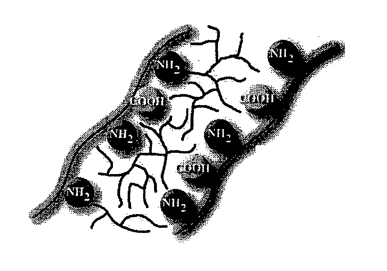
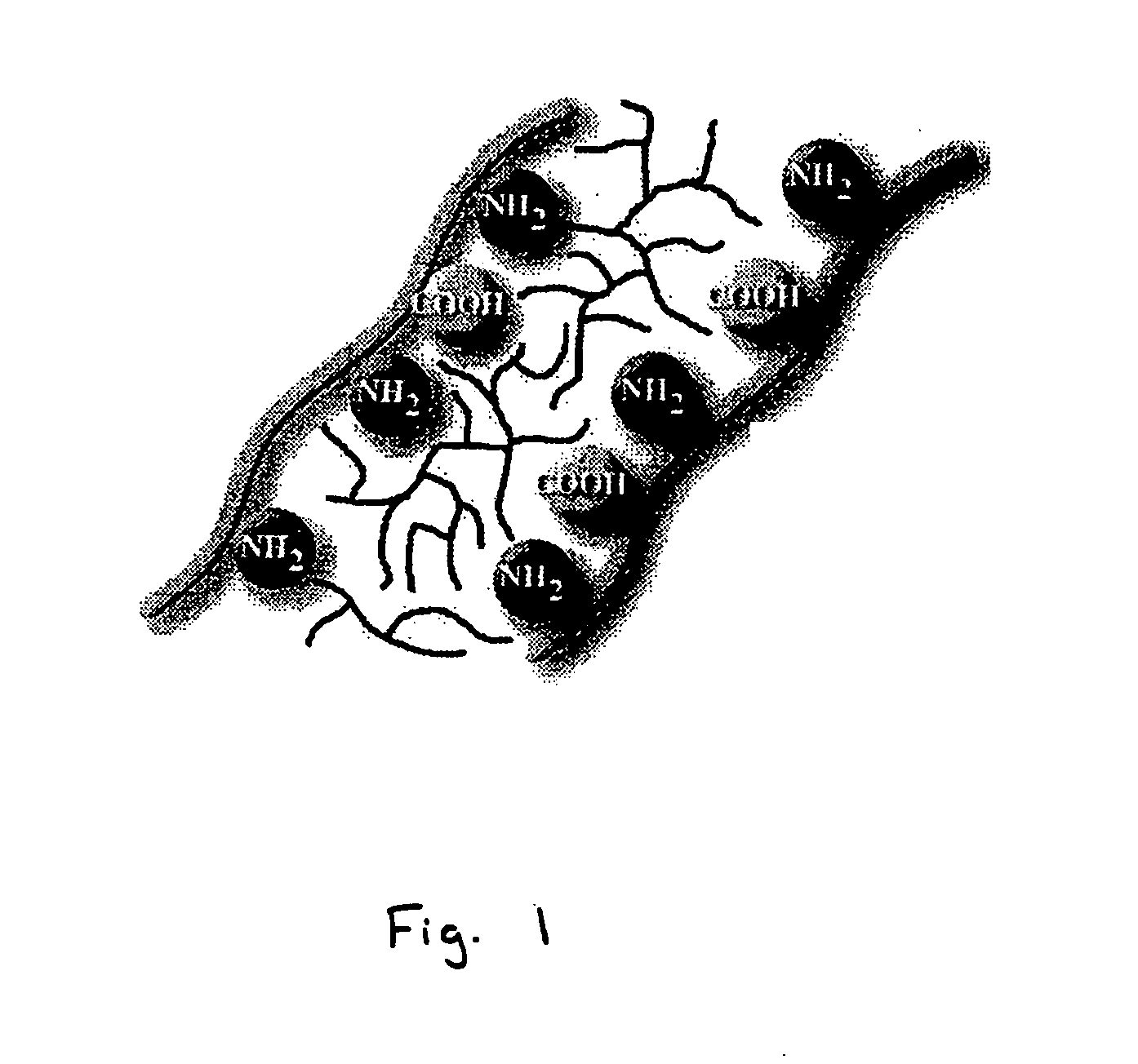
Bioprosthetic tissue preparation with synthetic hydrogels
a bioprosthetic tissue and hydrogel technology, applied in the field of medical devices, can solve the problems of ga fixed tissue being subject to calcification, eliciting severe immunological responses, and tissue enzymatically degraded, and achieve the effect of reducing tissue calcification and stiffness
- Summary
- Abstract
- Description
- Claims
- Application Information
AI Technical Summary
Benefits of technology
Problems solved by technology
Method used
Image

Examples
example 1
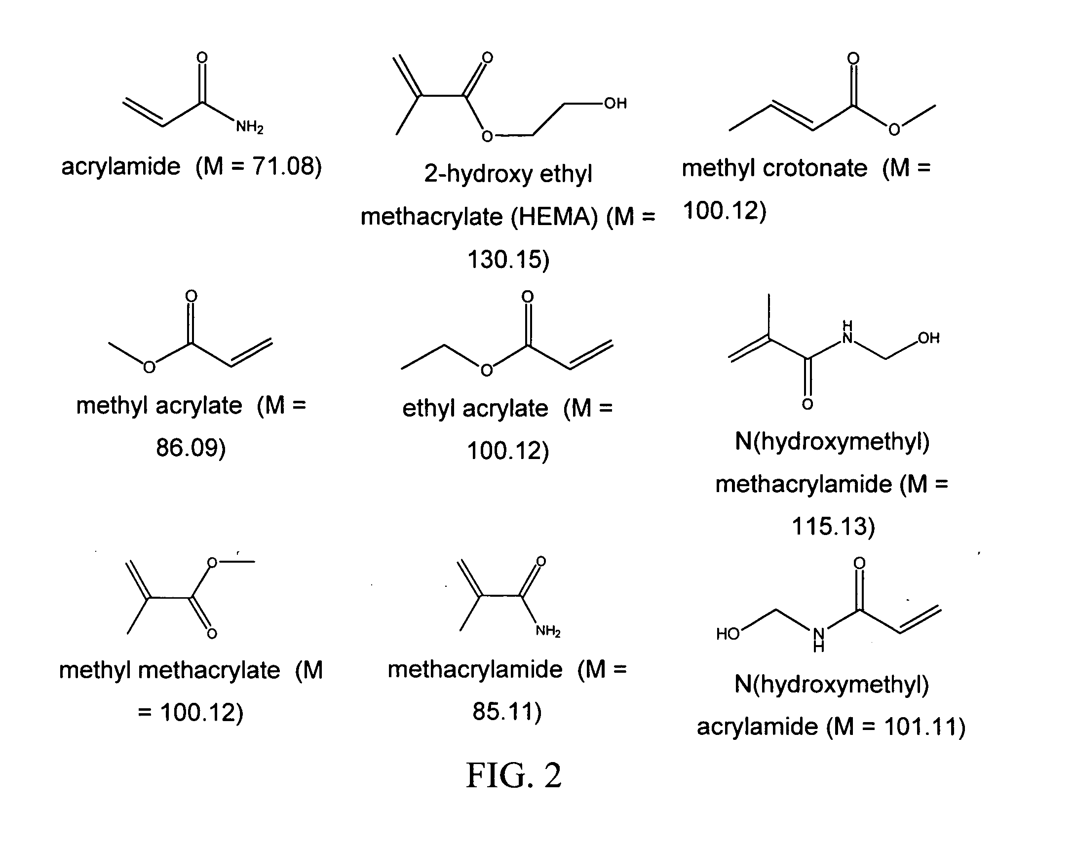
AAm+bAAm
[0088] Fresh porcine heart valve tissue was rinsed in a buffered saline solution. The tissue was subsequently removed from the buffered saline, and placed in a fresh buffered saline solution containing acrylamide (AAm; 30 g / 100 ml), N, N′-methylene bisacrylamide (bis-AAm: AAm ratio=1:36.5) and 2,2-dimethoxy-2-phenylacetophenone (DMPA; 0.4 mass % of total monomer) for 20 hours at 4° C. After removal of the tissue from the solution and removal of excess solution by blotting on tissue paper, the tissue samples were placed in a Petri dish, covered with fresh buffered saline, and exposed to long wave ultraviolet radiation (315-400 nm) for 20 minutes (10 minutes per side). Unreacted monomer was removed by 8×30 minute washes in buffered saline at 4° C. All solutions were sterilized by filtration prior to use.
[0089] In some embodiments, the monomer concentration is between 1 and 60 percent, preferably between 10 and 30 percent, by mass. The cross-linker concentration in some embod...
example 2
GA Control
[0090] Fresh, rinsed porcine tissue was cross linked by immersion in a buffered saline solution containing 0.2% glutaraldehyde (GA) at 4° C. for 7 days. This GA fixed tissue was subsequently processed according to the method outlined in Example 1.
example 3
HEMA+bAAm
[0091] Tissue was treated according to specifications in Example 1, with the exception that 20 g / 100 ml hydroxyethyl methacrylate (HEMA) was used instead of the 30 g / 100 ml acrylamide. The treatment ranges described with respect to Example 1 can be used.
PUM
 Abstract
Description
Claims
Application Information