Apparatus and method for marking tissue

a tissue and applicator technology, applied in the field of markers, can solve the problems of inability to define, affecting the exact recognition of the margins of the lesion, and affecting the visibility of the lesion by the imaging system

Inactive Publication Date: 2005-10-20
DEVICOR MEDICAL PROD
View PDF77 Cites 161 Cited by
  • Summary
  • Abstract
  • Description
  • Claims
  • Application Information

AI Technical Summary

Benefits of technology

[0035] An advantage of an injectable marker of the present invention is that such markers can be deployed using various surgical needles and various biopsy device sizes for placement at the biopsy site. This universal marker eliminates the need for multiple marker product specifically designed for each needle size. The injection marker type can be controlled injection related to the biopsy taken or physician preference.

Problems solved by technology

In the former case, a major problem is the ability to define the margins of the lesions at all times during the extraction process.
Visibility of the lesion by the imaging system may be hampered because of the distortion created by the extraction process itself as well as associated bleeding in the surrounding tissues.
Although the lesion is removed and all fluids are continuously aspirated from the extraction site, it is likely that the process will “cloud” the lesion, thus impairing exact recognition of its margins.
This makes it difficult to ensure that the entire lesion will be removed.
This is because such a lesion actually serves to mark or define the location of adjacent abnormal tissue, so the physician does not wish to remove the entire lesion and thereby lose a critical means for later re-locating the affected tissue.
However, oftentimes, either inadvertently or because the lesion is too small, the entire lesion is removed for evaluation, even though it is desired to remove only a portion.
Then, if subsequent analysis indicates the tissue to be malignant (malignant tissue requires removal, days or weeks later, of tissue around the immediate site of the original biopsy), it is difficult for the physician to determine the precise location of the lesion, in order to perform necessary additional procedures on adjacent potentially cancerous tissue.
While such a location system is effective, it is obviously intended and designed to be only temporary, and is removed once the surgery or other procedure has been completed.
Obviously, however, none of these approaches are useful for marking and delineating internal tissue abnormalities, such as lesions or tumors.
Of course, this method also is only a temporary means for marking the abnormal tissue, since eventually the drug will clear from even the abnormal tissue.
Additionally, once the abnormal tissue has been destroyed during treatment, the marker is destroyed as well.
However, while such staining techniques can be effective, it is difficult to precisely localize the stain.
Also, the stains are difficult to detect fluoroscopically and may not always be permanent.
Because such hydrogels undergo a relatively small amount of swelling and are not absorbable, so that the sterilization is not reversible, the devices described in the foregoing reference have found limited utility.

Method used


Image

  • Apparatus and method for marking tissue
  • Apparatus and method for marking tissue
  • Apparatus and method for marking tissue

Examples


Embodiment Construction

[0047] Before the present device and methods for modulation of appetite and satiety are described, it is to be understood that this invention is not limited to the specific methodology, devices. It is also to be understood that the terminology used herein is for the purpose of describing particular embodiments only, and is not intended to limit the scope of the present invention which will be limited only by the appended claims.

[0048] It must be noted that as used herein and in the appended claims, the singular forms “a”, “and”, and “the” include plural referents unless the context clearly dictates otherwise. Thus, for example, reference to “an active agent delivery system” includes a plurality of such devices and reference to “the method of delivery” includes reference to equivalent steps and methods known to those skilled in the art, and so forth.

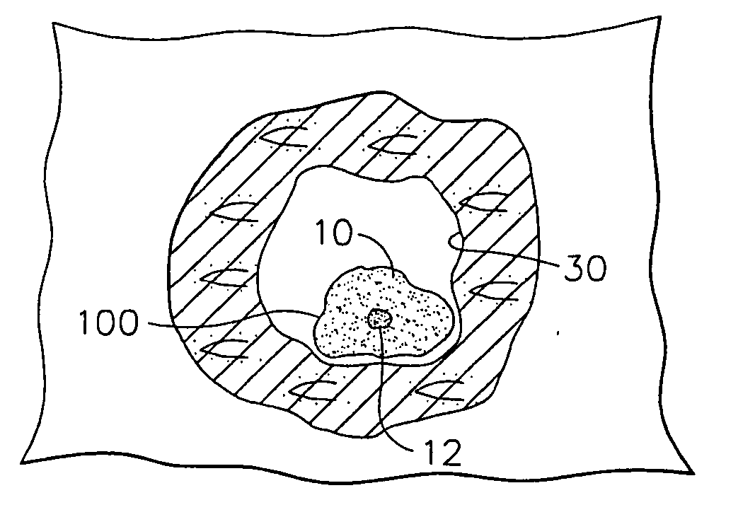
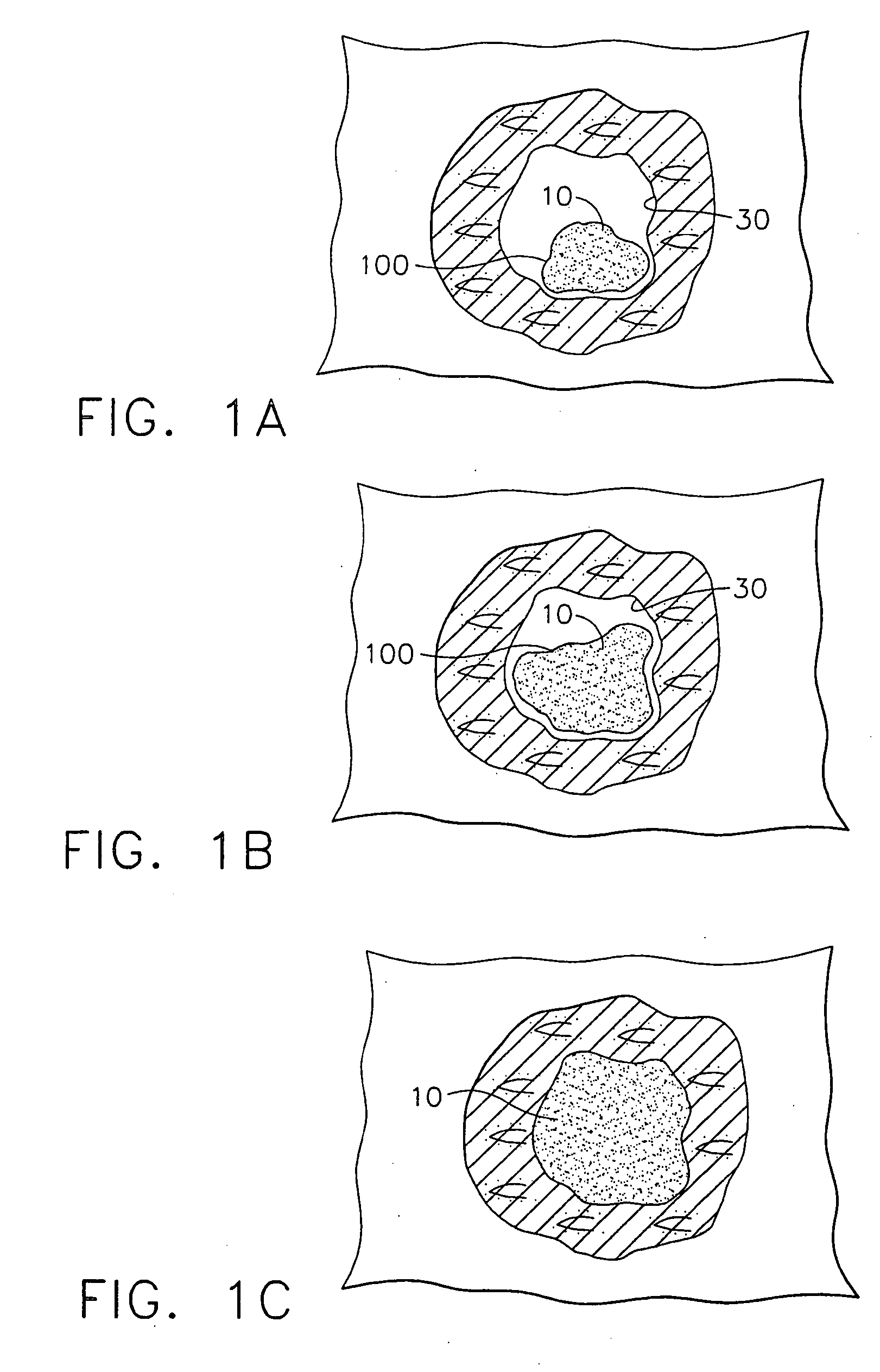
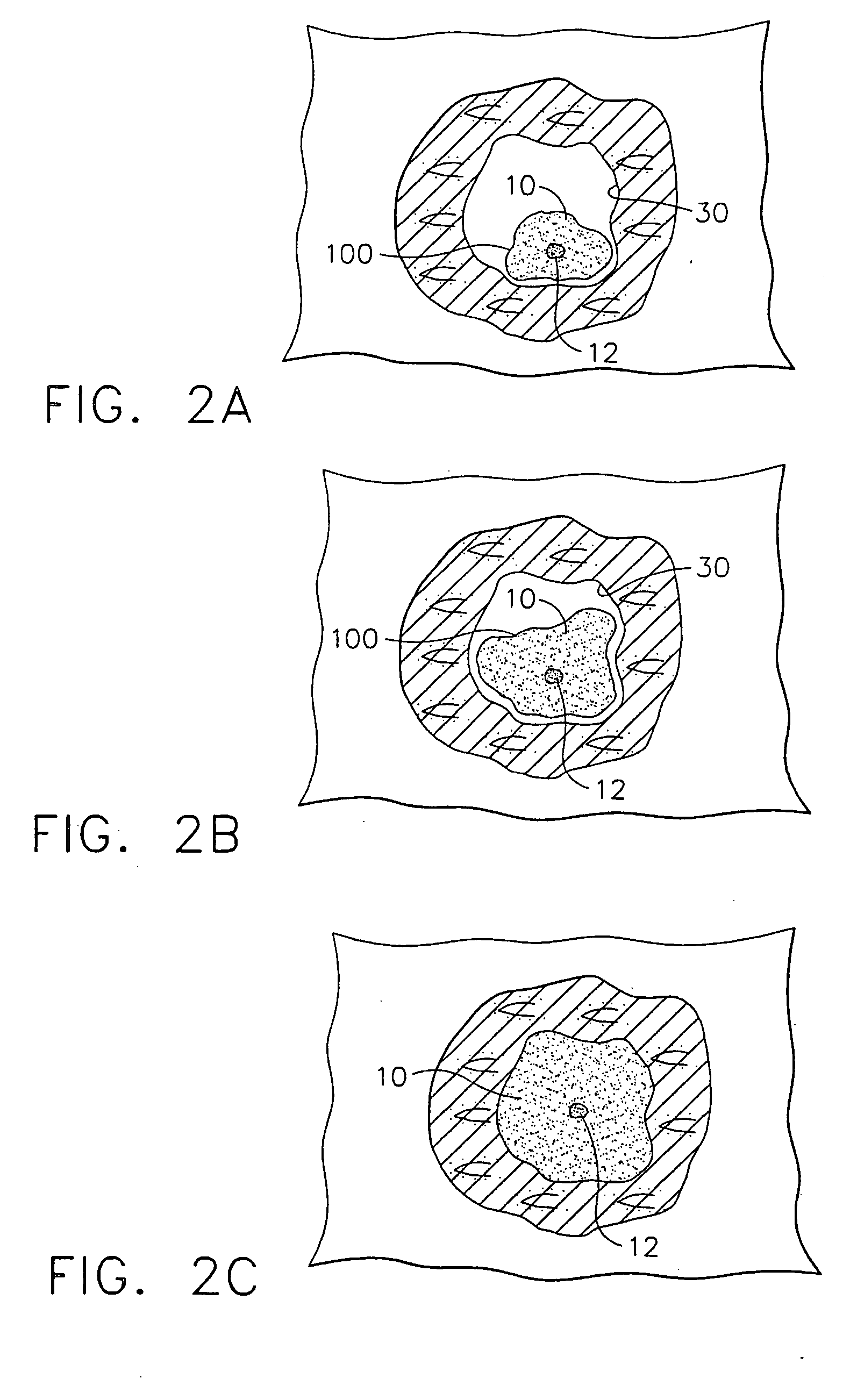
[0049] The invention features devices and methods for making and using a permanent implant marker that is detectable by at least two i...


PUM

PropertyMeasurementUnit
densityaaaaaaaaaa
densityaaaaaaaaaa
diameteraaaaaaaaaa

Abstract

The present invention includes methods and materials for implantable devices (markers) which are disclosed for permanently marking the location of a biopsy or surgery for the purpose of identification. The devices are remotely delivered, preferably percutaneously. Visualization of the markers is readily accomplished using various state of the art imaging systems. Preferred visualization is through MRI, X-ray and ultrasound. The markers function to provide evidence of the location of the lesion after the procedure is complete for reference during future examinations or procedures.

Description

FIELD OF THE INVENTION [0001] The present invention relates to markers to be employed at biopsy sites to permanently mark the site, and to methods and apparatus for applying the permanent marker. More particularly, the present invention relates to a marker that is optimally adapted for marking biopsy sites in human breast tissue with permanently placed markers that are detectable by MRI, ultrasound and X-ray. This invention relates to methods and devices for marking and defining particular locations in body tissue, particularly human tissue, and more particularly relates to methods and devices for permanently defining the location and margins of lesions detected in biopsy cavity walls. BACKGROUND OF THE INVENTION [0002] In the U.S. alone approximately one million women will have breast biopsies because of irregular mammograms and palpable abnormalities. Biopsies can include surgical excisional biopsies and stereotactic and ultrasound guided needle breast biopsies. In the case of ima...

Claims


Application Information

Patent Timeline
Patent Type & Authority Applications(United States)
IPC IPC(8): A61B10/00A61B19/00A61B10/02A61L31/18
CPCA61B10/02A61L31/18A61B2019/5408A61B19/54A61B90/39A61B2090/3908
Inventor BECKMAN, ANDREW T.ONWUMERE, FIDELISLUDZACK, MICHAEL ROBERTSAMPLES, CHARLES ROBERTBARBUR, ANA
Owner DEVICOR MEDICAL PROD
Why Eureka
Try Eureka