

Peptide capable of binding to nanographite structures
a nanographite and peptide technology, applied in the direction of peptide/protein ingredients, instruments, drug compositions, etc., can solve the problems of difficult to use nanographite structures such as carbon nanohoms and carbon nanotubes
- Summary
- Abstract
- Description
- Claims
- Application Information
AI Technical Summary
Problems solved by technology
Method used
Image

Examples
example 1
[0040] The surface of carbon in a form of a sinterd round bar in ambient gas pressure of 6×10⁴ Pa of Ar gas was ablated with high-power CO2 gas laser beam (output power 100 W, pulse width 20 ms, continuous wave), and the resultant soot-like substance was suspended in ethanol, then ultrasonic agitation (frequency 40 kHz, 60 minutes) and decantation were repeated 4 times to obtain single-wall carbon nanohoms. About 200 mg of the single-wall carbon nanohoms was put into 40 ml of nitric acid at a concentration of about 70%, and reflux was conducted for 1 hour at 130° C. After the reflux, the resultant was neutralized and washed by repeating dilution with ion-exchange water, centrifugation, and disposal of the supernatant, and water-soluble single-wall carbon nanohorns having a functional group (including a carboxyl group) were prepared.
[0041] 1.2 mg of nitric acid-treated carbon nanohoms was suspended in 2 ml of 0.1 M 2-morpholinoethanesulfonic acid monohydrate (hereinafter referred to...
example 2
[0056] Single-wall carbon nanotube synthesized by chemical vapor deposition, Hipco (Carbon Nanotechnologies Inc., Texas), was treated with 1×10⁻⁵ Torr for 5 hours at 1750° C., and then reflux was conducted for 30 minutes at about 130° C. in nitric acid at a concentration of about 70%. After that, neutralization with sodium hydroxide and washing with distilled water were conducted, and single-wall carbon nanotubes having a functional group (including a carboxyl group) were prepared.
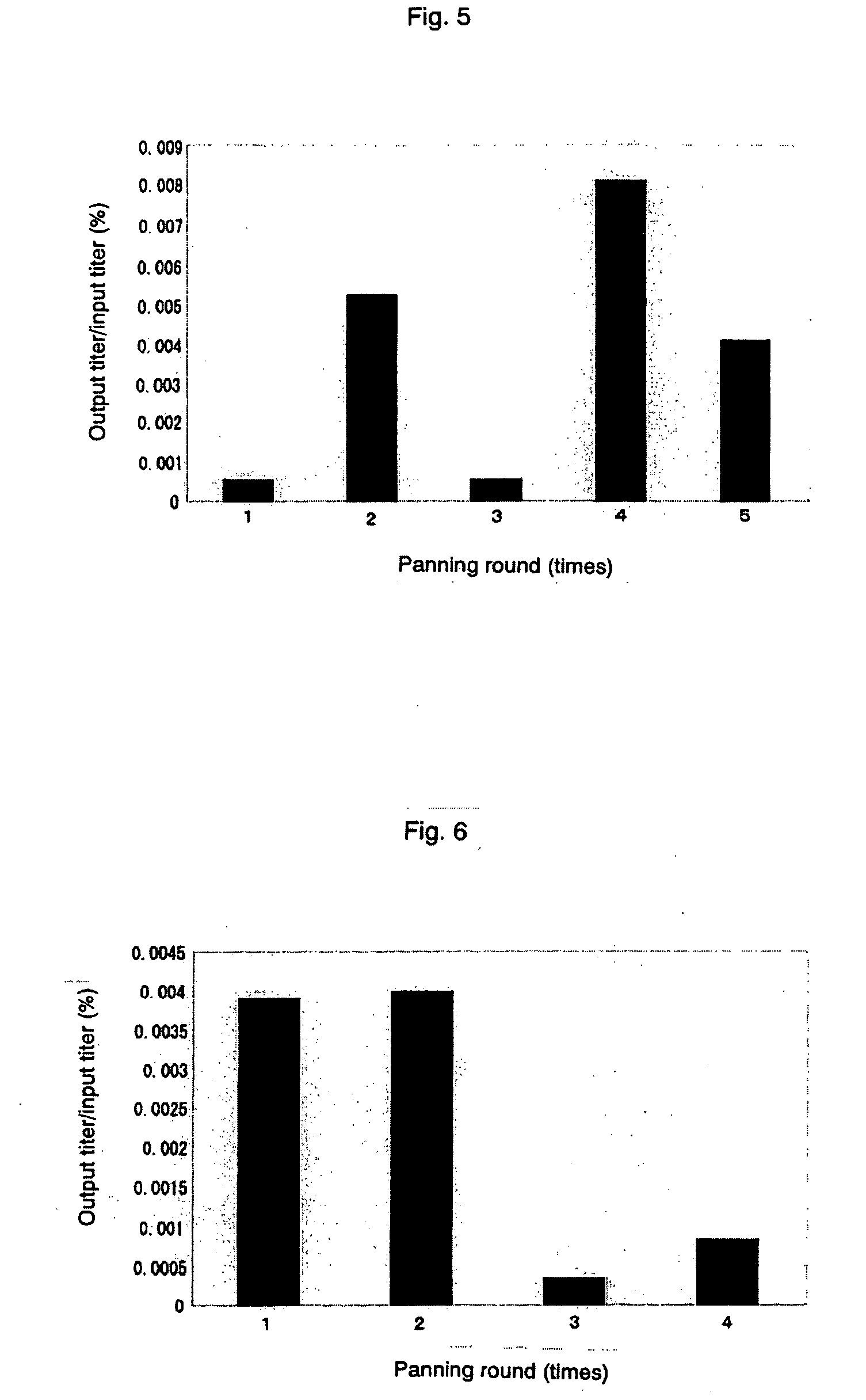
[0057] The nitric acid-treated single-wall carbon nanotubes thus obtained were biotinylated and solid-phased on streptavidin-coated magnetic beads in a same manner as shown in Example 1. With the use of the solid-phased carbon nanotubes, panning experiment was conducted with D12 library and C7C library in a same manner as shown in Example 1, with the proviso that for C7C library, input titer of phage at the 1st round was set at 2.4×10¹¹, and after that, it was set at 1.2×10¹⁰ as in Example 1.
[0058] The c...
example 3
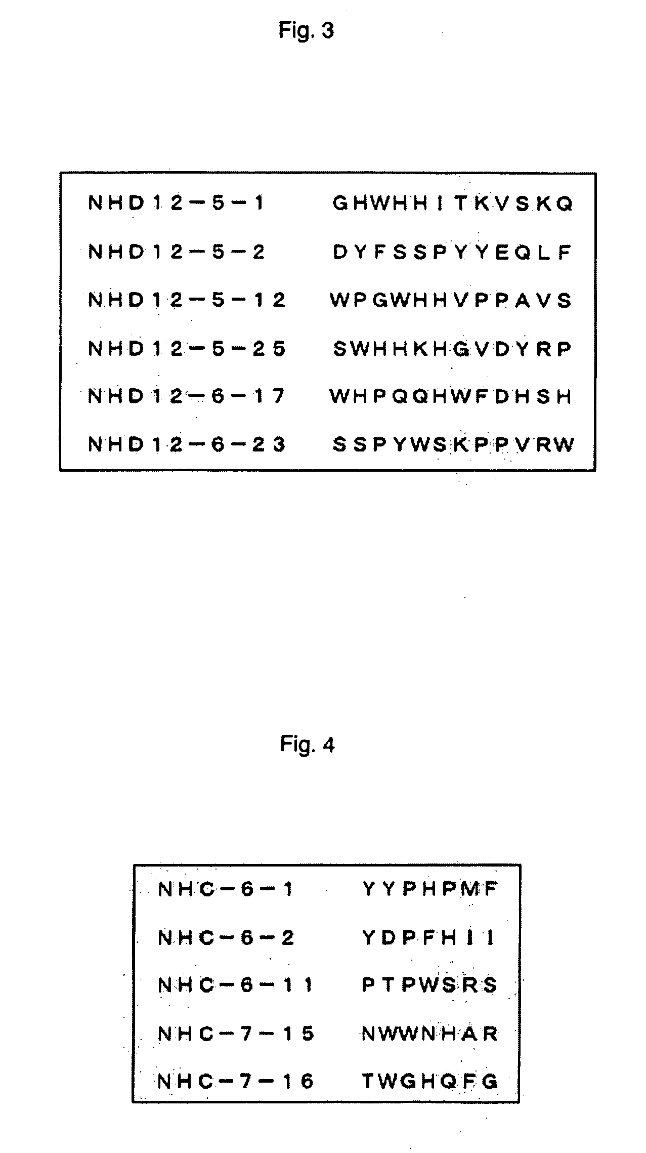
[0059] With regard to a suspension of single-wall carbon nanotubes, unlike that of carbon nanohoms, it is possible to separate only carbon nanotubes by centrifugation. In the same manner as shown in Example 1, panning operations to untreated single-wall carbon nanotubes were conducted with the use of D12 library. However, prepanning operation was conducted with a 96-well ELISA plate (Immulon 4HBX, Dynex Technologies, Inc., VA), binding reaction of phages was set to 2 hours, and separation of single-wall carbon nanotubes after the reaction was conducted by putting the reaction liquid into a 1.8 ml plastic tube and centrifuging it for 8 minutes at 15000 rpm using the high-speed microcentrifuge (mentioned above). Further, input titer of phage was set at 2.7×10¹¹ in the 1st to 2nd rounds, and 2.7×10¹⁰ in the rounds after that. The changes in the ratio of input titer and output titer are shown in FIG. 9. Some of displayed peptide sequences expected from the determined base sequences are ...
PUM
| Property | Measurement | Unit |
|---|---|---|
| Thickness | aaaaa | aaaaa |
| Structure | aaaaa | aaaaa |
| Size | aaaaa | aaaaa |
Abstract
Description
Claims
Application Information