
Cell-permeable enzyme activation reporter that can be loaded in a high throughput and gentle manner

a reporter and cell-permeable technology, applied in the field of cell-permeable enzyme activation reporter, can solve the problems of hampered cellular mechanism study and diagnosis, unable to meet the intimidating terminology, and the introduction of these molecules is not without significant cos

Inactive Publication Date: 2005-12-22
RGT UNIV OF CALIFORNIA
View PDF0 Cites 8 Cited by
  • Summary
  • Abstract
  • Description
  • Claims
  • Application Information

AI Technical Summary

Problems solved by technology

2001), this same fortification presents challenges to those exogenous molecules that may exert beneficial effects, such as cell membrane-impermeant pharmaceuticals.
Furthermore, the study of cellular mechanisms and diagnosing diseases and disorders are also hampered.
The introduction of these molecules, however, have not been without significant cost, both to the target cells as well as to the proper understanding of data resulting from these molecular perturbations.
The disadvantages of these methods match the intimidating terminology.
The primary disadvantage of each is that the target cells are severely stressed, resulting in aberrant activation of intracellular proteins.
In all of these methods but pinocytosis, physical holes are poked into the cell membranes, disrupting membrane integrity.
Microinjection and optoporation have the added disadvantages in that they can only be performed on only one cell (or few cells) at a time, severely limiting their usefulness.
These methods also require expensive, dedicated equipment, and in many cases, large amounts of the membrane-impermeant molecules.
These in vitro methods typically deliver the nucleic acid molecules into only a fraction of the total cell population, and they tend to damage large numbers of cells.
However, these techniques have, to date, shown limited usefulness for in vivo cellular delivery.
Moreover, even with cells in vitro, such methods are of extremely limited usefulness for delivery of proteins.
Given the high levels of circulating folate in vivo, the usefulness of this system has not been fully demonstrated.
In both of these approaches, however, the confounding effects of the membrane breaching-enabling molecules cannot be discounted once the cargo molecules have entered the cell, although Rothbard et al.
However, these studies may be confounded by the observation that due to TAT's high affinity for the plasma membrane, TAT conjugates bind to cell membranes even at low temperatures and remain after washing.
While TAT can breach the cell membrane barrier, too much TAT (concentrations greater than 5 μM) often kills cells.
Unfortunately, toxic concentrations are necessary to introduce sufficient concentrations of some reporter molecules, such as fluorescently-tagged TAT (Vives, Brodin et al.
Once in the cell, however, the cargo no longer requires TAT's assistance; however, its continued linkage to the cargo may ultimately inhibit the cargo's intended activity (Stein, Weiss et al.

Method used


Image

  • Cell-permeable enzyme activation reporter that can be loaded in a high throughput and gentle manner
  • Cell-permeable enzyme activation reporter that can be loaded in a high throughput and gentle manner
  • Cell-permeable enzyme activation reporter that can be loaded in a high throughput and gentle manner

Examples


example 1

Characterization of TAT(49-57) Conjugated to a Substrate Peptide

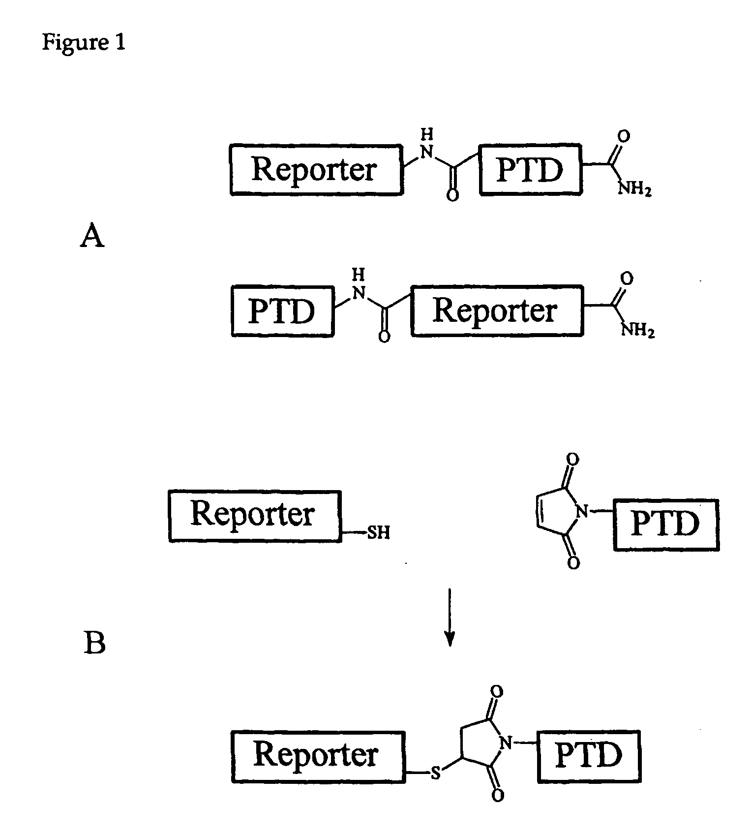
[0125] To determine whether a kinase substrate linked to TAT(49-57) was accessible to cytosolic enzymes, rat basophilic leukemia (RBL) cells were incubated with 1 μM calcium-calmodulin dependent kinase II peptide substrate (F-CamKII; SEQ ID NO:11) conjugated to TAT(49-57)(SEQ ID NO:2) through a peptide bond (F-CamKII-TAT; SEQ ID NO:12). To determine if F-CamKII-TAT (SEQ ID NO:12) is phosphorylated by its target kinase, CamKII, cells were loaded with F-CamKII-TAT and then their contents analyzed for phosphorylated F-CamKII using the Cell Activity by Capillary Electrophoresis (CACE) (also known as LMS or Laser Micropipet System, see Methods, below). With the CACE, a single cell can be rapidly lysed and the cellular contents loaded with nearly 100% efficiency into a capillary exceptionally quickly (33×10−3 seconds) (Sims, Meredith et al. 1998; Meredith, Sims et al. 2000; Li, Sims et al. 2001). Together with cell lysis, el...

example 2

Characterization of Substrate Peptides Conjugated to TAT(49-57) by a Disulfide Bond

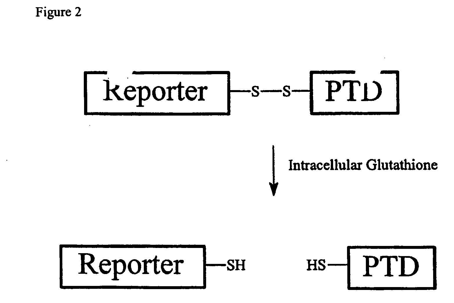
[0129] Previous investigators developed cleavable PTD-cargo conjugates by engineering a disulfide linkage between the two domains (Stein, Weiss et al. 1999; Hallbrink, Floren et al. 2001). In the oxidative extracellular environment, the disulfide bond remained intact, but in the reductive intracellular environment, the disulfide bond quickly reduced releasing the cargo from the PTD (Meister 1989; Hallbrink, Floren et al. 2001). TAT(49-57)(SEQ ID NO:2) was conjugated via a disulfide bond to cysteines added to substrate peptides for CamKII (F-CamKII-C; SEQ ID NO:13) or PKB (F-PKB-C; SEQ ID NO:14), denoted F-CamKII-C-SS-TAT (SEQ ID NO:15) and F-PKB-C-SS-TAT (SEQ ID NO:16), respectively. The TAT(49-57)-Conjugated substrates were incubated with cells (HT1080 or HT1080 / PTEN cells) and the contents of the cells were analyzed by the CACE. HT1080 cells have constitutive PKB activity due to a mutated allele of...

example 3

Phosphorylation of Substrate Peptides Delivered by a Disulfide-Linked TAT(49-57)

[0132] To determine whether the detached peptide substrates were in the cytosol and accessible to kinases, the intracellular phosphorylation of the translocated cargo was studied. In the initial experiment, F-CamKII-C-SS-TAT (SEQ ID NO:15) at 750 nM extracellular concentration was loaded into HT1080 cells. The cells were incubated with ionomycin (500 nM) for 15 min to increase the intracellular concentration of free Ca2+ and activate the CamKII enzyme (Worrell and Frizzell 1991). The electrophoretic traces from these cells displayed a second peak in addition to the peak of due to the non-phosphorylated F-CamKII-C. The migration time of the second peak was identical to that of phosphorylated F-CamKII-C loaded into cells. 52+3% (mean±SEM) (n=5) of the F-CamKII-C was phosphorylated as determined by comparison of the peak areas. At least half of the F-CamKII-C was in the cytosol and accessible to the kinase...


PUM

PropertyMeasurementUnit
Fluorescenceaaaaaaaaaa
Chemiluminescenceaaaaaaaaaa
Phosphorescence quantum yieldaaaaaaaaaa

Abstract

A membrane traversing peptide conjugate comprises (i) a label, (ii) a target peptide, attached to the label, and (iii) a transduction domain, attached to the target peptide. The target peptide is a reactant for a chemical reaction occurring in a cell.

Description

[0001] This application claims the benefit of U.S. Provisional Application No. 60 / 530,875 filed 17 Dec. 2003.STATEMENT OF ACKNOWLEDGMENT OF GOVERNMENT SUPPORT [0002] This invention was made with Government support under Grant Nos. CA91216, GM57015 and NS39310, awarded by the National Institutes of Health. The Government has certain rights in this invention.BACKGROUND [0003] The body prevents a number of defenses against outside invaders, which manifest in tissues, organs and systems. The skin sets up a passive first line of defense, for example, while the immune system actively assails those invaders that have breached other barriers. But even cells themselves—regardless of their type—erect a barrier which grants them control over cellular entry: the cell membrane. For the most part, only by duping cell-surface molecules or by physical disruption may invaders (e.g., molecules, viruses, bacteria) gain entry. [0004] While cell membranes form a desirable barrier that prevents the flux ...

Claims


Application Information

Patent Timeline
IPC IPC(8): C07K14/47C12Q1/48
CPCC12Q1/485C07K14/47
Inventor ALLBRITTON, NANCYSIMS, CHRISTOPHERROSSI, FRANCISSOUGHAYER, JOSEPH
Owner RGT UNIV OF CALIFORNIA
Learn More