One-step enzymatic and amine detection technique

a technology of detection method, which is applied in the field of one-step enzymatic and amine detection method, can solve the problems of inability to detect a single molecule,

Inactive Publication Date: 2006-01-05
KIMBERLY-CLARK WORLDWIDE INC
View PDF99 Cites 32 Cited by
  • Summary
  • Abstract
  • Description
  • Claims
  • Application Information

AI Technical Summary

Problems solved by technology

However, conventional techniques for confirming the presence of “clue cells” are often complicated and slow.
Unfortunately, such tests are undesired in that they require performance by a professional and utilize caustic chemicals.
Unfortunately, the techniques described above su

Method used


Image

  • One-step enzymatic and amine detection technique
  • One-step enzymatic and amine detection technique
  • One-step enzymatic and amine detection technique

Examples


example 1

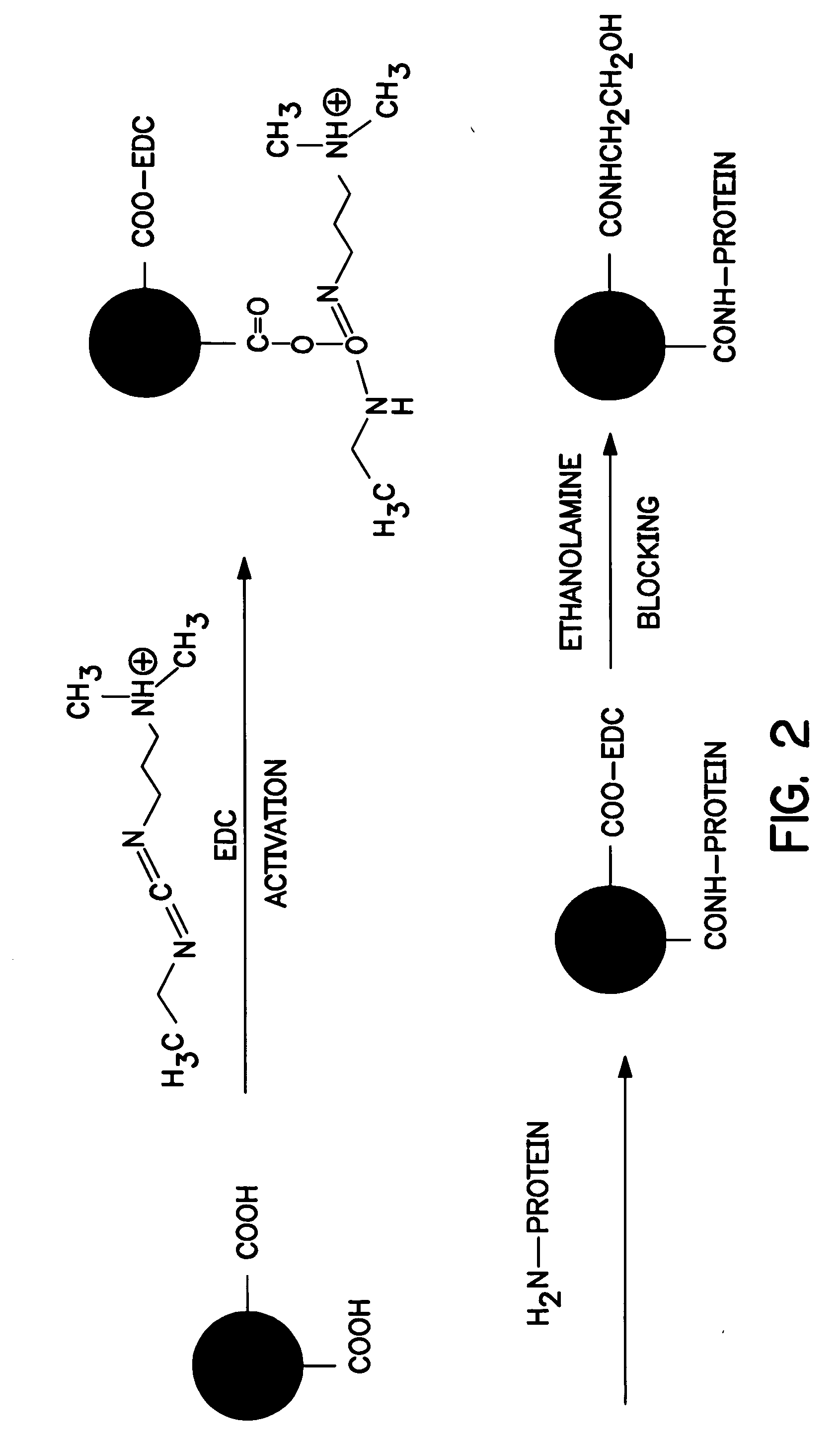
[0094]β-casein was initially conjugated to dyed particles. Specifically, 2 milliliters of blue carboxylated particles (0.3-micrometer particle size, Bangs Laboratories, Inc. of Fisher, Ind.) were washed once with phosphate-buffered saline (PBS from Polysciences, Inc. of Warrington, Pa.) and then suspended in 1 milliliter of PBS. 36 milligrams of carbodiimide (Polysciences, Inc.) in 1 milliliter of PBS was added and the mixture was shaken for 30 minutes. The particles were washed twice with a borate buffer (Polysciences, Inc.), and then suspended in 1 milliliter of borate buffer. 1 milligram of β-casein (Sigma-Aldrich Chemical Co., Inc. of St. Louis, Mo.) was added and the mixture was shaken overnight at room temperature. The particles were washed once with the borate buffer and then re-suspended in 500 microliters of borate buffer. 1 milliliter of ethanolamine solution (0.1 molar, Polysciences Inc.) was added to the particles and shaken for 30 minutes. The particles were then washed...

example 2

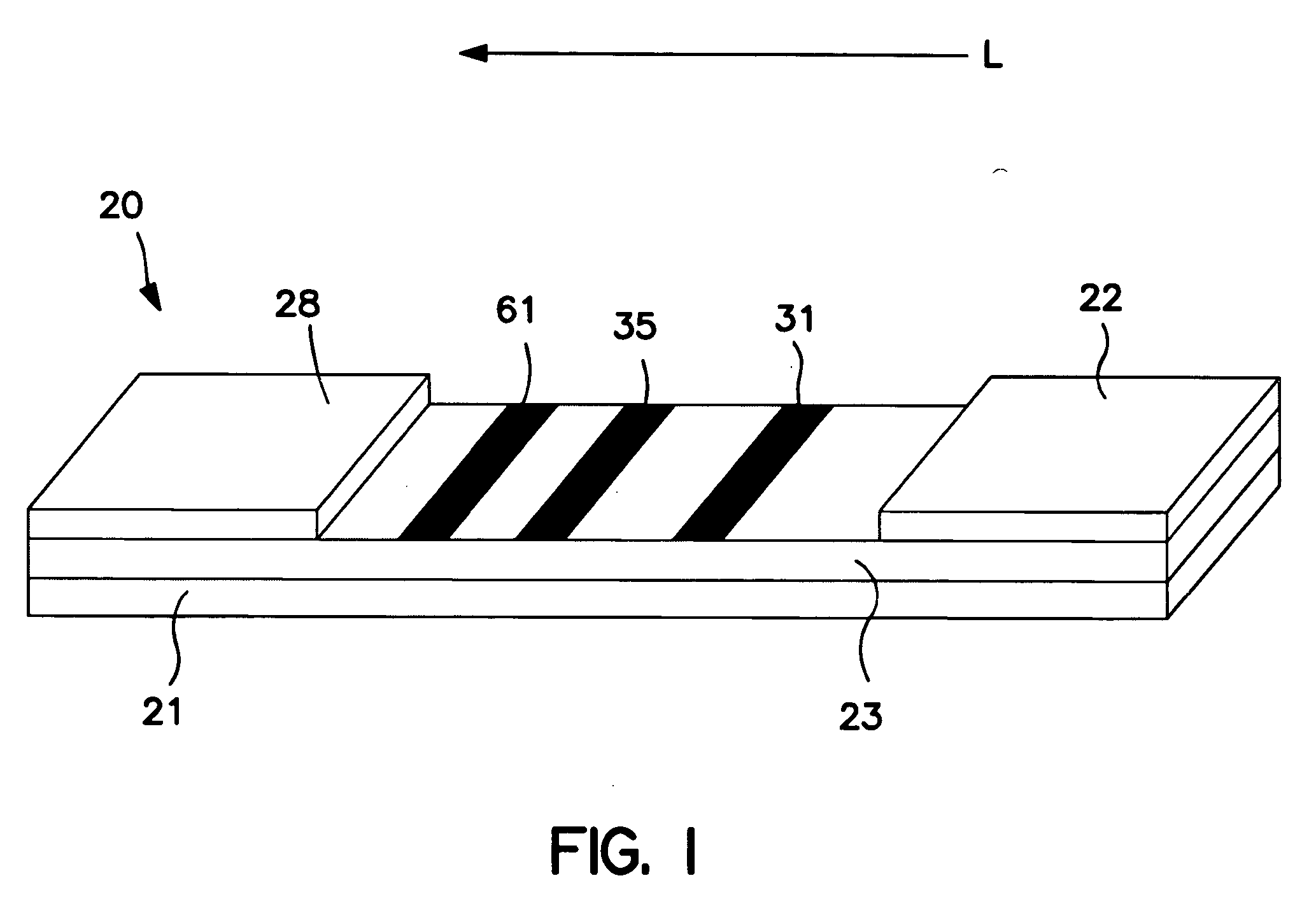
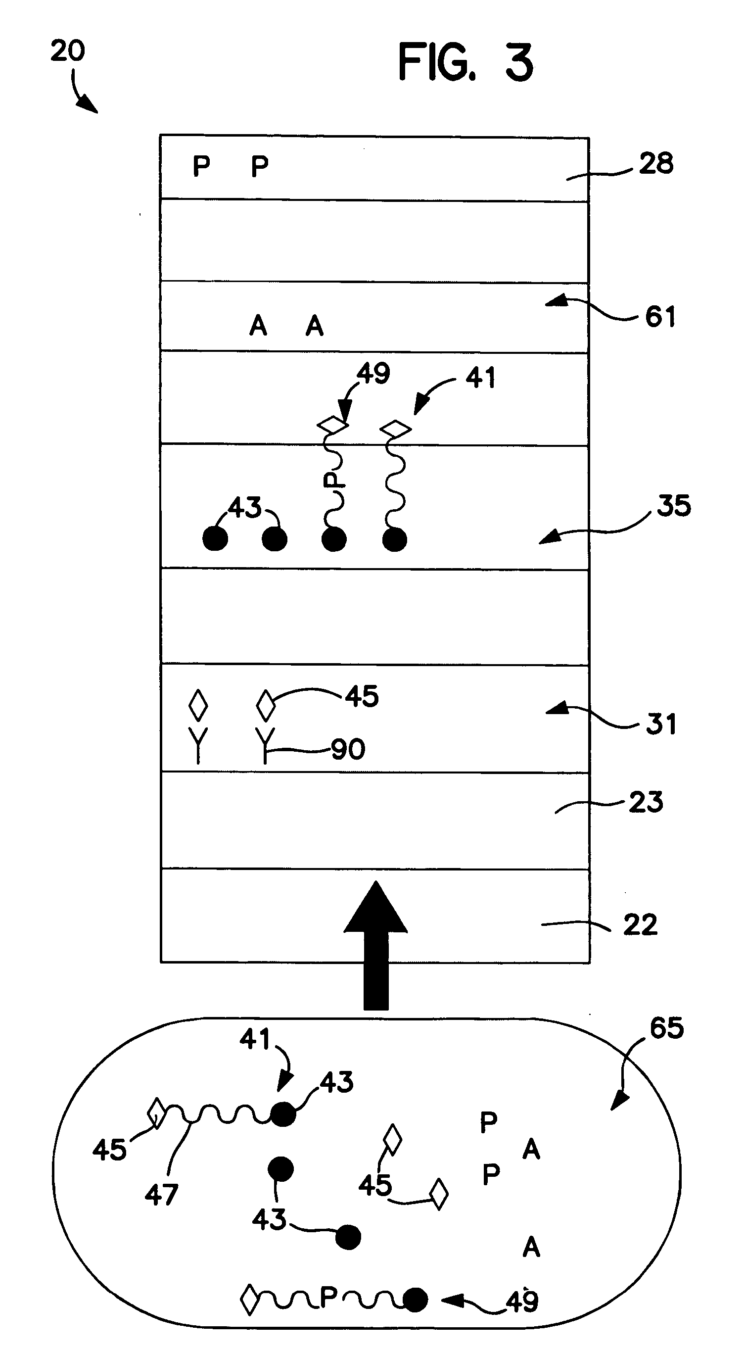
[0096] The ability to form a membrane-based device for amine and / or enzyme assays was demonstrated. Initially, Millipore HF12002 porous nitrocellulose membranes were laminated onto corresponding supporting cards having a length of approximately 30 centimeters. Streptavidin (1.0 milligram per milliliter, Sigma-Aldrich Chemical Co., Inc.) was striped onto the membrane to form a first enzyme detection zone and Goldline™ (a polylysine solution obtained from British Biocell International) was striped onto the membrane (downstream from the first enzyme detection zone) to form a second enzyme detection zone. Alpha-naphtholbenzein (ANB) (5 milligrams per milliliter, Sigma-Aldrich Chemical Co., Inc.) was also striped onto the membrane (downstream from the enzyme detection zones) to form an amine detection zone. The membrane was dried for 1 hour at 37° C. A cellulosic fiber wicking pad (Millipore Co.) was attached to the end of the membrane closest to the amine detection zone. The assembled c...

example 3

[0097] The ability to form a membrane-based device for amine and / or enzyme assays was demonstrated. Initially, Millipore HF12002 porous nitrocellulose membranes were laminated onto corresponding supporting cards having a length of approximately 30 centimeters. Anti-biotin antibody (2.0 milligrams per milliliter, Sigma-Aldrich Chemical Co., Inc.) was striped onto the membrane to form a first enzyme detection zone and Goldline™ (a polylysine solution obtained from British Biocell International) was striped onto the membrane (downstream from the first enzyme detection zone) to form a second enzyme detection zone. ANB dye (Sigma-Aldrich Chemical Co., Inc.) was also striped onto the membrane (downstream from the enzyme detection zones) to form an amine detection zone. The membrane was dried for 1 hour at 37° C. A cellulosic fiber wicking pad (Millipore Co.) was attached to the end of the membrane closest to the ANB dye zone. The assembled card was then cut into 4-millimeter wide devices....


PUM

PropertyMeasurementUnit
Coloraaaaaaaaaa
Magnetismaaaaaaaaaa
Affinityaaaaaaaaaa

Abstract

A technique for detecting the presence or quantity of an enzyme (or enzyme inhibitor) and/or an amine within a test sample is provided. For example, in one embodiment, a diagnostic test kit is employed that utilizes reactive complexes that each includes a substrate joined (e.g., covalently bonded, physically adsorbed, etc.) to a reporter and a separation species. Upon contacting the reactive complexes, enzymes may cleave the substrate and release the reporter. Moreover, the test kit may also employ a chemichromic dye, i.e., a dye that exhibits a detectable color change upon chemical reaction with one or more functional groups, such as amino groups. The signal generated (directly or indirectly) by the reporter and chemichromic dye may then be used to indicate the presence or quantity of an enzyme (or enzyme inhibitor) and amine, respectively, within the test sample.

Description

BACKGROUND OF THE INVENTION [0001] The rapid diagnosis of infection is becoming increasingly important to improving the effectiveness of subsequent treatment. Vaginal infection (“vaginitis”), for example, exists in three primary forms, i.e., bacterial vaginosis, candidal vaginitis (“yeast”), and trichomonas vaginitis (“trich”). Various techniques have been developed in an attempt to rapidly diagnose individual forms of vaginitis. Bacterial vaginosis, for example, has been diagnosed using “clue cells” (vaginal epithelial cells with adherent surface bacteria). However, conventional techniques for confirming the presence of “clue cells” are often complicated and slow. Techniques have been utilized that detect an elevated pH level in an infected sample. [0002] Bacterial vaginosis and trichomonas vaginitis (primarily caused by the protozoan, Trichomonas vaginalis) may also cause a “fishy” odor that stems from an elevated level of amines, such as putrescine (1,4-diaminobutane), cadaverine...

Claims


Application Information

Patent Timeline
IPC IPC(8): C12Q1/68G01N33/573G01N33/53C12Q1/04G01N33/558
CPCG01N33/558C12Q1/04
Inventor SONG, XUEDONGBOGA, RAMESHBABUCHIDEBELU-EZE, CHIBUEZE OBI
Owner KIMBERLY-CLARK WORLDWIDE INC
Why Eureka
Try Eureka