Integration of direct binding label-free biosensors with mass spectrometry for functional and structural characterization of molecules

a biosensor and mass spectrometry technology, applied in the field of integration of direct binding label-free biosensors with mass spectrometry for functional and structural characterization of molecules, can solve the problems of limited utility of previously known methods of combining detection assays and structural analysis

Inactive Publication Date: 2006-01-05
SRU BIOSYST
View PDF0 Cites 36 Cited by
  • Summary
  • Abstract
  • Description
  • Claims
  • Application Information

AI Technical Summary

Benefits of technology

[0004] One embodiment of the invention provides a method of analyzing or identifying one or more molecules. The method comprises contacting a sample comprising the one or more molecules with a colorimetric resonant reflectance optical sensor such that one or more of the one or more molecules become immobilized to the colorimetric resonant reflectance optical sensor. The immobilized one or more molecules are eluted from the colorimetric resonant reflectance optical sensor and subjected to mass spectrometry analysis. The one or more molecules immobilized to the colorimetric resonant reflectance optical sensor can be detected. The one or more molecules immobilized to the colorimetric resonant reflectance optical sensor can be directly detected by a shift in peak wavelength value (PWV). The one or more molecules immobilized to the colorimetric resonant reflectance optical sensor can be detected using a label. A peak wavelength value (PWV) signal can also be detected. The detecting can comprise use of an indicator molecule of equal, greater, or lesser molecular mass of the one or more molecules immobilized to the colorimetric resonant reflectance optical sensor. The one or more molecules can be quantified. The one or more molecules can be immobilized to the colorimetric resonant reflectance optical sensor by one or more moieties on the surface of the colorimetric resonant reflectance optical sensor. The one or more moieties can be TiO, RaM Fc, avidin, biotin, an antibody, an antibody fragment, a nucleic acid molecule, protein A, hybrids of protein A, protein G, hybrids of protein G, protein L, hybrids of protein L, high density PVA, CHO or a combination thereof. The colorimetric resonant reflectance optical sensor can be coupled to a flow sys...

Problems solved by technology

Previously known methods of combining detection assays with structural analysis are extremely limited in utility by the subfemtomole quantities of bound materia...

Method used


Image

  • Integration of direct binding label-free biosensors with mass spectrometry for functional and structural characterization of molecules
  • Integration of direct binding label-free biosensors with mass spectrometry for functional and structural characterization of molecules
  • Integration of direct binding label-free biosensors with mass spectrometry for functional and structural characterization of molecules

Examples


example 1

Tandem BIND / MALDI-MS Experiment

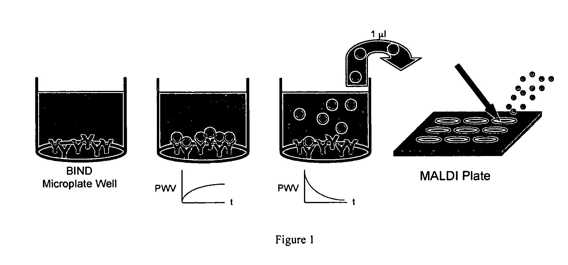
[0062] Experiments were performed to show that material bound to a colorimetric resonant reflectance optical sensor surface can be efficiently eluted and analyzed by mass spectrometry. A capture antibody was adsorbed to a colorimetric resonant reflectance optical sensor surface. The corresponding antigen was allowed to bind to the antibody during application of the sample on the colorimetric resonant reflectance optical sensor. Any material bound specifically to the antibody was subsequently eluted from the surface. An aliquot of the eluted material was then applied for mass spectrometry analysis. The eluted material was mixed with the appropriate MALDI matrix and added to a MALDI plate and used for (TOF) MS analyses.

[0063]FIG. 1 shows the protocol used to combine the colorimetric resonant reflectance optical sensor technique with a MALDI type mass spectroscopy experiment. The process comprises adsorb antibodies on a colorimetric resonant reflectanc...

example 2

Matrix Assisted Laser Desorption Ionization (MALDI) Mass Spectroscopy (MS)

[0064] A colorimetric resonant reflectance optical sensor TiO sensor was pre-rinsed with PBS 3 times and left at room temperature for 30 min. A baseline reading was taken for a few min and 10 ul of 1 mg / ml of human IgG or chicken IgY was diluted into 90 ul PBS already in the well for a final concentration of the antibodies of 100 ug / mL. The protein was put into the well and allowed to bind to the TiO surface for 90 min. Unbound protein solution was removed from the well and the wells were rinsed 3 times each with 200 ul of PBS.

[0065] Another baseline reading was taken for a few minutes and then 1 ul of 1 mg / ml of anti-human IgG (Fab)2 was put into the wells, which were coated with either hIgG (Red) or cIgY (Yellow) and allowed to incubate for 60 min. All the unbound protein solution was removed from the wells and the wells were rinsed 3 times with PBS. The binding signal was monitored for stability for few ...

example 3

Tandem BIND / MALDI-MS Experiment—MS Data

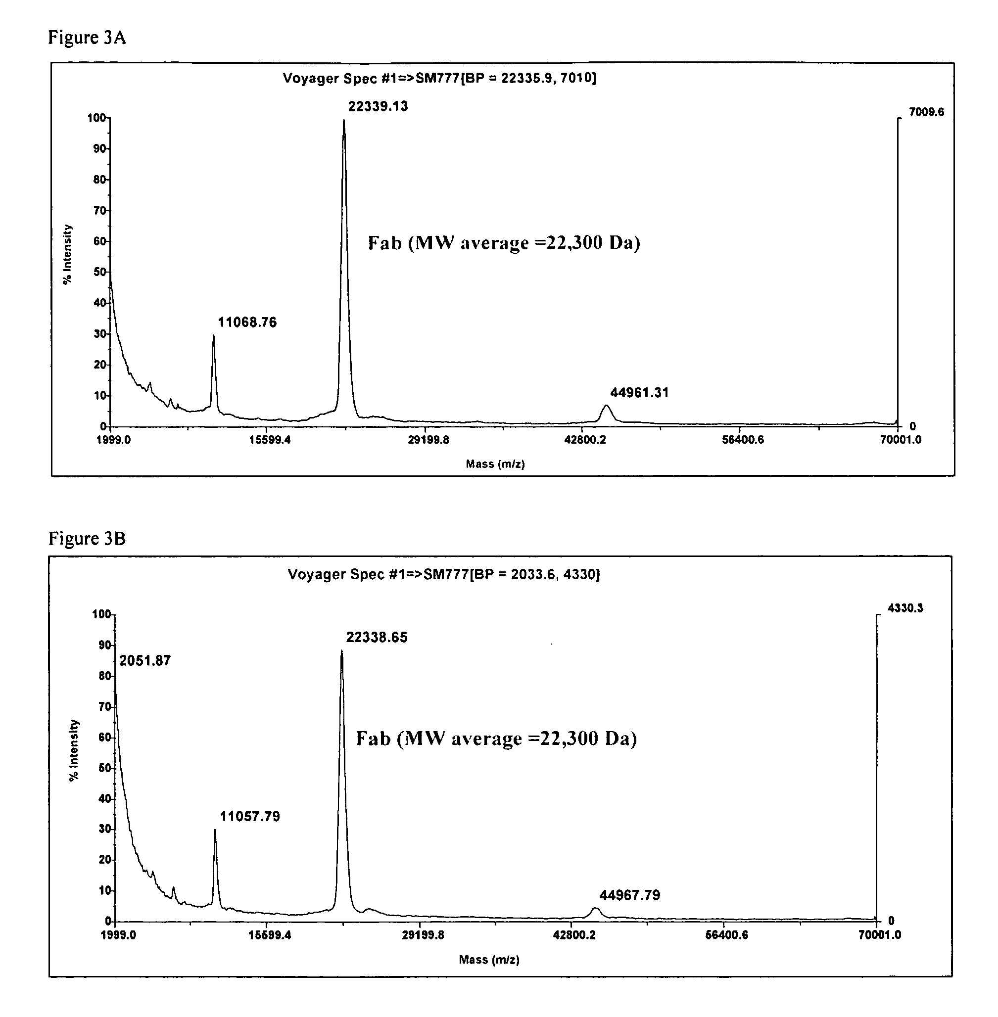
[0067]FIG. 3A shows the data from a control solution containing the Fab that was applied to the MS prior to exposure to the sensor surface. The primary peak is at 22300, the other two peaks are signature peaks related to the parent molecular mass

[0068]FIG. 3B shows the MALDI-MS data from the solution that was eluted from the sensor surface. The mass spectra is identical to the control spectra shown in FIG. 3A.


PUM

PropertyMeasurementUnit
Massaaaaaaaaaa
Wavelengthaaaaaaaaaa
Binding constantaaaaaaaaaa

Abstract

The invention provides methods for the detection, quantification, identification and structural analysis of one or more molecules. Mass spectrometry (MS) is not a universal detector as all molecules do not ionize equally well leading to poor signal to quantity information. MS can be optimized to identify the specific mass of a binding component when the presence of a material is known. Colorimetric resonant reflectance optical sensors provide a universal mass detector in that nearly all biological masses give equally proportional signals. The combined methods allow selection and or detection with quantification of all masses binding to the sensor with the ability to identify specific molecules by their individual masses and structure analyses.

Description

PRIORITY [0001] This application claims the benefit of U.S. application Ser. No. 60 / 583,560, filed on Jun. 28, 2004, which is incorporate herein by reference in its entirety.BACKGROUND OF THE INVENTION [0002] Detection assays combined with structural analysis can provide complementary information on function and structure of molecules. Previous work has demonstrated the utility of this combined approach for applications such as ligand fishing, epitope mapping, and amino acid sequencing, but only in the low throughput sample-limited context of a microfluidic channel-surface plasmon resonance (SPR)-based systems. See, Nelson et al., BIA / MS of Epitope-Tagged Peptides Directly from E. coli Lysate: Multiplex Detection and Protein Identification at Low-Femtomole to Subfemtomole Levels. Analytical Chemistry 1999, 71:2858-2865; Nelson et al., Biosensor chip mass spectrometry: A chip-based proteomics approach. Electrophoresis 2000, 21:1155-1163. Previously known methods of combining detectio...

Claims


Application Information

Patent Timeline
IPC IPC(8): C12Q1/68G01N33/53
CPCG01N21/27G01N33/542G01N33/6848G01N33/54373G01N33/543
Inventor LI, PETERLIN, BOWILLIAMS, CHRISLAING, LANCE
Owner SRU BIOSYST
Why Eureka
Try Eureka