Assay methods for state-dependent calcium channel agonists/antagonists
- Summary
- Abstract
- Description
- Claims
- Application Information
AI Technical Summary
Benefits of technology
Problems solved by technology
Method used
Image
Examples
example 1
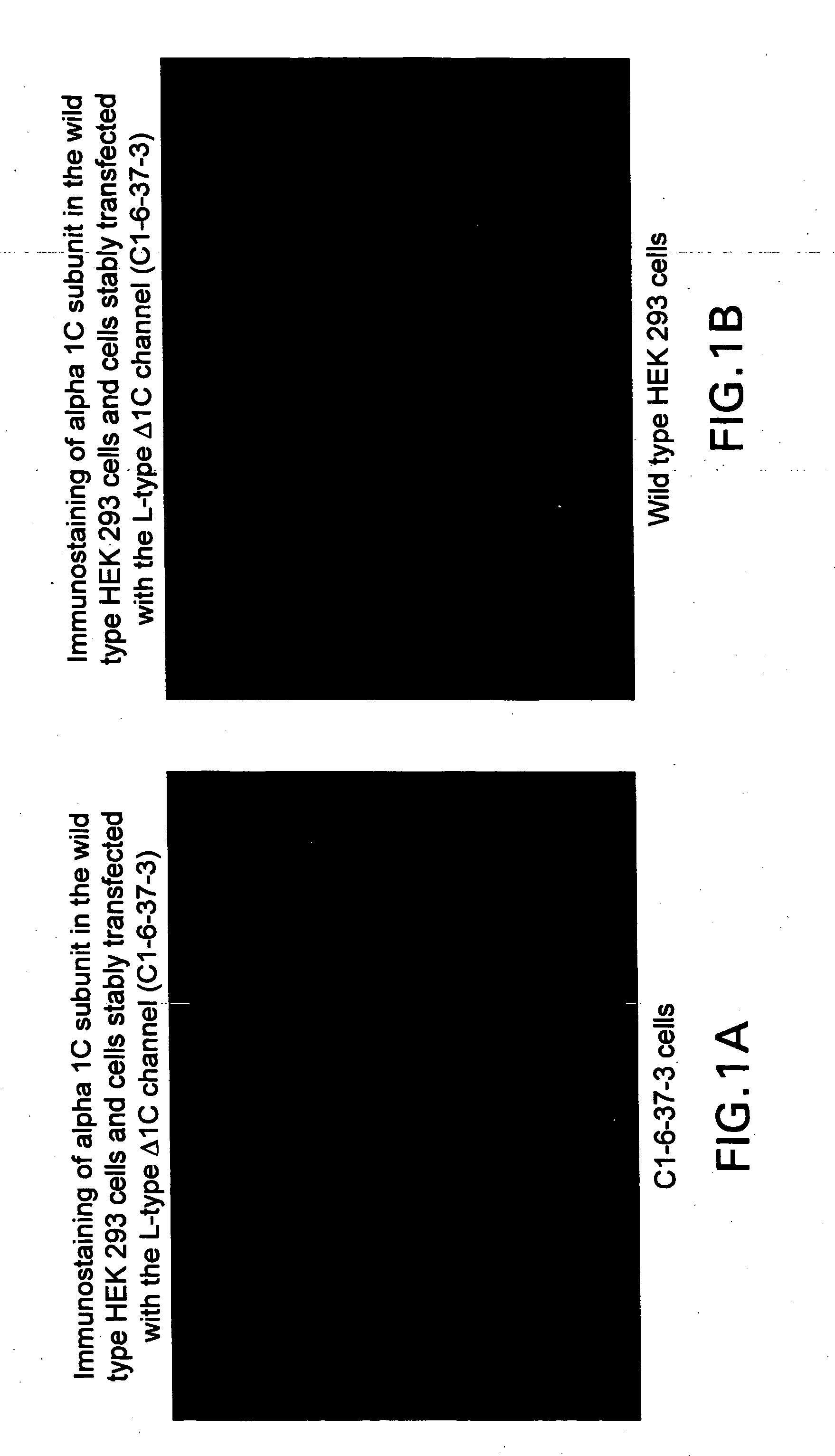
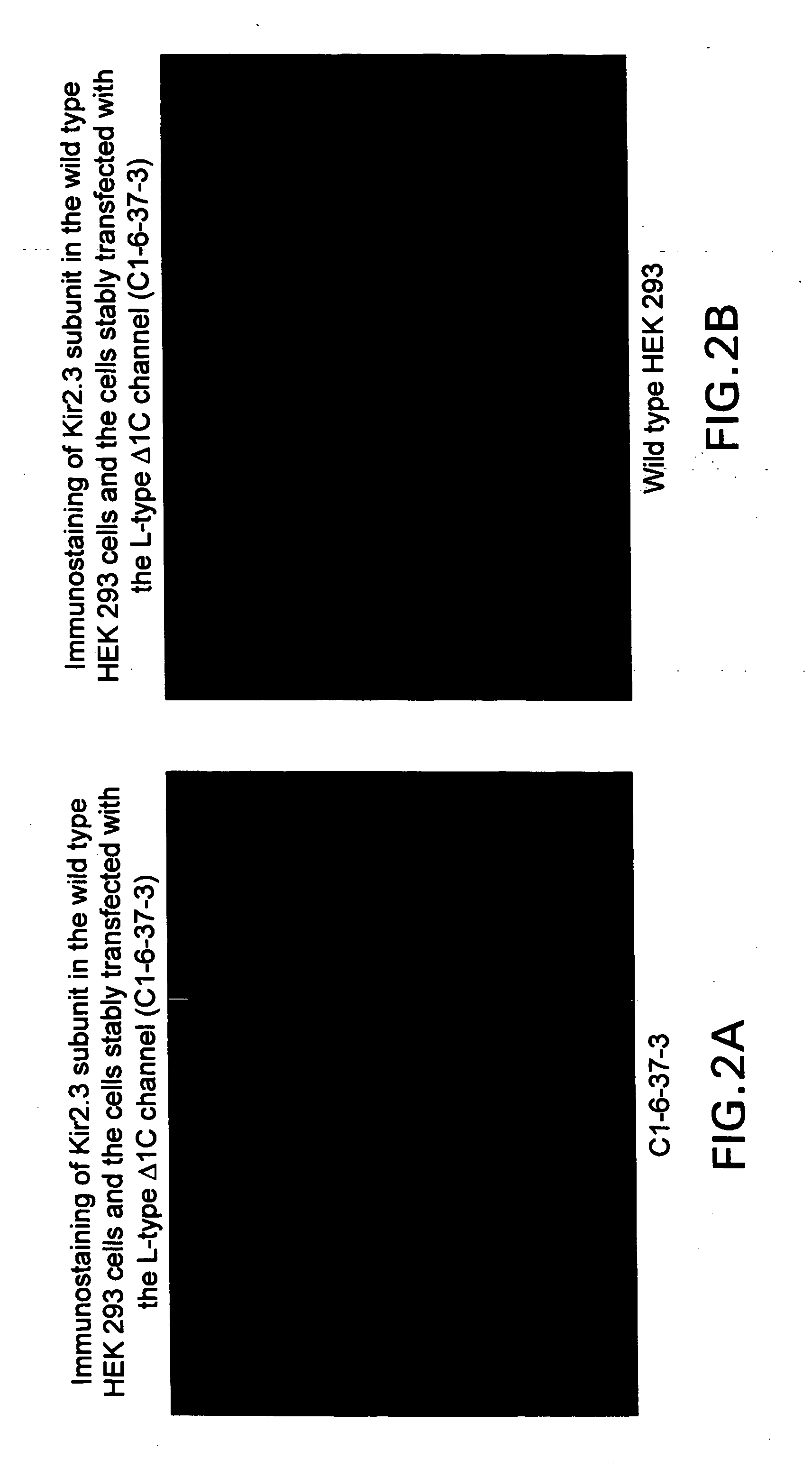
[0089] Immunofluorescence staining was all performed at room temperature. Cells were washed three times with Dulbecco's phosphate buffered saline (D-PBS) and then fixed with 4% paraformldehyde for 30 min. After three washes with D-PBS, the cells were blocked and permeabilized with TBS (10 mM Tris-HCl, pH 7.5, and 150 mM NaCl) containing 4% nonfat dry milk and 0.1% Triton X-100 for 1 hr, and incubated with the affinity purified polyclonal antibodies against human alpha 1C or kir2.3 for 1 hr. Then the cells were washed three times with TBS and incubated with the secondary antibody (Cy3-conjugated anti-rabbit IgG, at 1:250, Jackson ImmunoResearch, PA) for 1 hr. The cells were finally washed with D-PBS three times and viewed under indirect immunofluorescence on a Zeiss Axioskop microscope. FIGS. 1 and 2 show that cells were successfully transfected and expressing calcium and potassium channels, respectively, on their plasma membranes.
example 2
[0090] Hek 293 cells were stably transfected with the alpha 1C subunit of the L-type Calcium ion channel and Kir 2.3 inward K+ rectifying channel (C1-6-37-3 cells). Calcium influx into the cells was measured in a FLIPR™ (Molecular Devices, CA). The C1-6-37-3 cells were seeded into black 96 well plates with clear bottoms coated with poly-D-lysine at density of 50000 cells / well and cultured overnight. Next day the cells were washed twice with assay buffer containing 137 mM NaCl; 0.34 mM Na2HPO4; 4.2 mM NaHCO3; 0.44 mM KH2PO4; 0.41 mM MgSO4; 0.49 mM MgCl2; 20 mM HEPES; 5.5 mM D-glucose and 0.1% BSA and incubated with Fluo-3AM (final concentration 4 μM, Molecular probe) for 1 hr at 37° C., 5% CO2 and 95% O2. After cells were washed four times either with resting condition (5.8 K+) or depolarized condition (30 mM K+), the cell plate was placed into the FLIPR™ to monitor cell fluorescence (λEX=488 nm, λEM=540 nm) before and after the addition of calcium blockers and agonists (final 85.8 m...
example 3
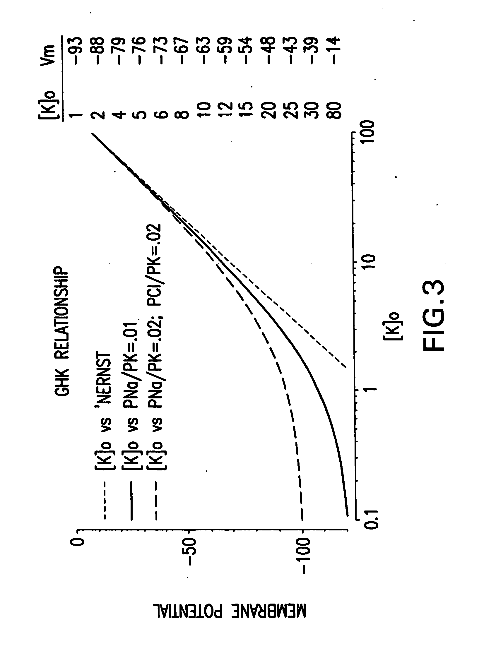
[0095] Cellular membrane potentials were measured using an Axopatch 200B patch amplifier (Axon Instruments Inc., Foster City, Calif.) in current clamp more using the ‘perforated patch’ clamp method (Horn and Korn). The patch pipette contained (in mM): 120 KMeSO4, 20 KCl, 9 Mg2Cl, 10 HEPES, Nystatin 200 μg / ml, pH7.3. The bath solution contained (in mM): 140 NaCl, 1.2 Mg2Cl, 10 HEPES, 1.3 Ca2Cl, 21 D-glucose, pH7.4. Standard electrophysiological methods were employed. Changes in extracellular potassium were made by additions of a concentrated stock to the standard bath solution to the appropriate dilution. FIG. 3 shows the relationship between extracellular potassium ([K]o) and cell membrane potential. Three situations are shown. One is the prediction of the Nernst equation for a perfectly K-selective membrane. The other curves show the effects of partial permeability by other ions, Na+ and / or Cl−. Membrane potential can be set in a non-voltage clamped cell by adjusting external potas...
PUM
| Property | Measurement | Unit |
|---|---|---|
| Time | aaaaa | aaaaa |
| Electric charge | aaaaa | aaaaa |
| Volume | aaaaa | aaaaa |
Abstract
Description
Claims
Application Information
 - R&D
- Intellectual Property
- Life Sciences
- Materials
- Tech Scout
- Unparalleled Data Quality
- Higher Quality Content
- 60% Fewer Hallucinations
Browse by: Latest US Patents, China's latest patents, Technical Efficacy Thesaurus, Application Domain, Technology Topic, Popular Technical Reports.
© 2025 PatSnap. All rights reserved.Legal|Privacy policy|Modern Slavery Act Transparency Statement|Sitemap|About US| Contact US: help@patsnap.com


