Nuclear transfer embryo formation method

a technology of nuclear transfer and embryos, applied in the field of nuclear transfer embryo formation methods, can solve the problems of inherently invasive and clearly damaging the spatial organization of cytoplasts, inability to enucleate cytoplasts, and inability to fully enucleate cytoplasts, so as to improve the yield of competent embryos

Inactive Publication Date: 2006-01-19
TRUSTEES OF TUFTS COLLEGE
View PDF15 Cites 15 Cited by
  • Summary
  • Abstract
  • Description
  • Claims
  • Application Information

AI Technical Summary

Benefits of technology

[0017] The methods of the invention have advantages over methods of nuclear transfer that employ mechanical enucleation (e.g., enucleation using a micropipette to remove the nucleus) of the oocyte for several reasons. For example, using physical enucleation, the nucleus along with other important spindle associated elements are also removed. These spindle associated elements include, for example, centrosome factors (α-tubulin, pericentrin), kinases (e.g., C-mos, MPF, PAPK) and competence factors (e.g., cdc25C, spindlin). Several of these elements are important in forming a nuclear transfer embryo that is developmentally competent (e.g., embryo's ability or likelihood to develop successfully in vivo following an embryo transfer and implantation to a recipient mammal). The method of the invention removes the endogenous nucleus from the oocyte, while allowing several of these spindle associated elements to remain in the oocyte, thereby potentially significantly improving yield of competent embryos.

Problems solved by technology

However, physical enucleation is generally technically demanding, time consuming, inherently invasive and clearly damaging to cytoplast spatial organization.
Moreover, in certain instances, development of reconstructed embryos is inherently inefficient.
However, certain studies have reported that exposure of metaphase I and MII oocytes to etoposide, a topoisomerase II inhibitor, and cycloheximide yields enucleated cytoplasts with limited ability to support cleavage or blastocyst development, and term development of reconstructed embryos has not been reported.

Method used


Image

  • Nuclear transfer embryo formation method
  • Nuclear transfer embryo formation method
  • Nuclear transfer embryo formation method

Examples


example 1

Improved Method of Cloning Through the Introduction of Donor Cell Nucleus Prior to Completion of Enucleation Process

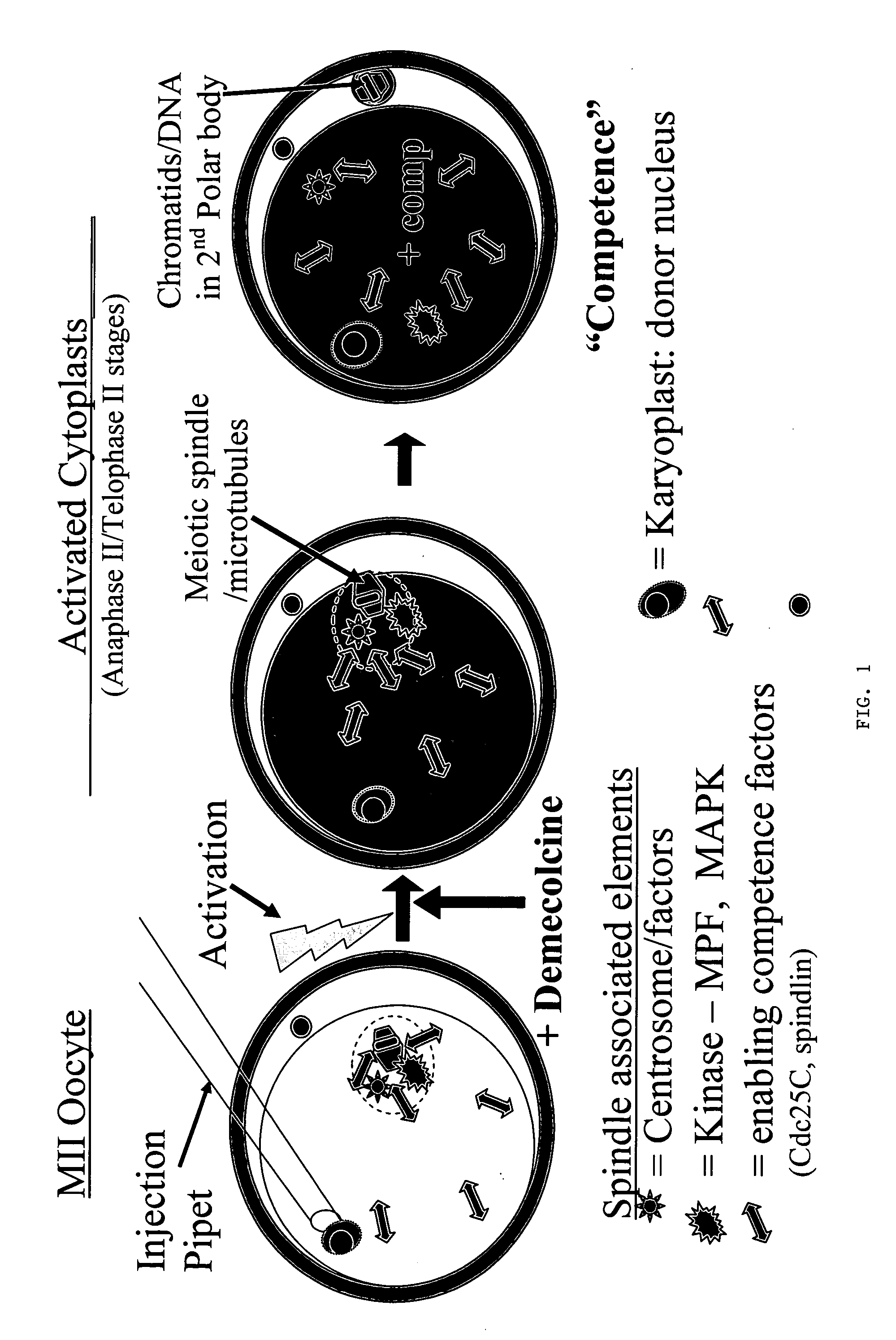
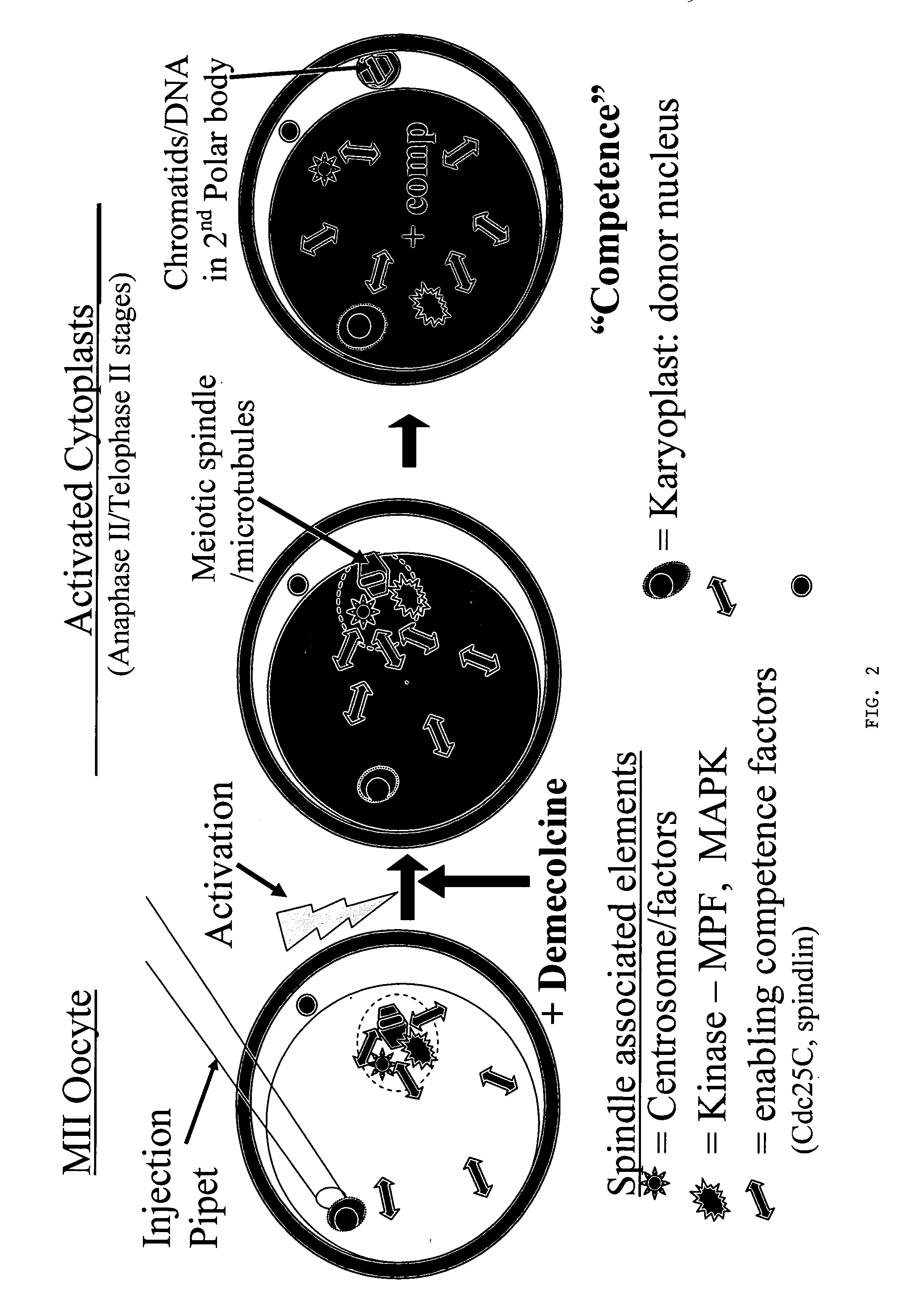
[0081] Progress has continued with experiments designed to compare in vitro development of control embryos (parthenotes) with that of reconstructed NT embryos prepared by conventional NT, telophase NT, IE and a novel simultaneous IE-NT paradigm (SIE / NT; see Table 1 below). As in all previous experiments, abattoir-sourced oocytes were obtained having been submitted to 26 h of maturation in ACM media @5% CO2 in air. Oocytes were denuded (enzyme / vortex) and activated (5 μM ionomyciin for 5 min, + / −CHX). See Example 2 for detail on methods. IE was performed by treatment of oocytes with demecolcine (0.4 μg / ml) starting at 1.5 h and ending 5 h post-activation (p.a.) and nuclei were injected 1.5-3 h p.a. In the case of SIE / NT, donor karyoplasts (fibroblasts) were injected immediately prior to or immediately after activation. In vitro embryo development was evaluated over a p...

example 2

Demecolcine-Induced Oocyte Enucleation for Somatic Cell Cloning: Coordination Between Cell Cycle Egress, Kinetics of Cortical Cytoskeletal Interactions, and Second Polar Body Extrusions

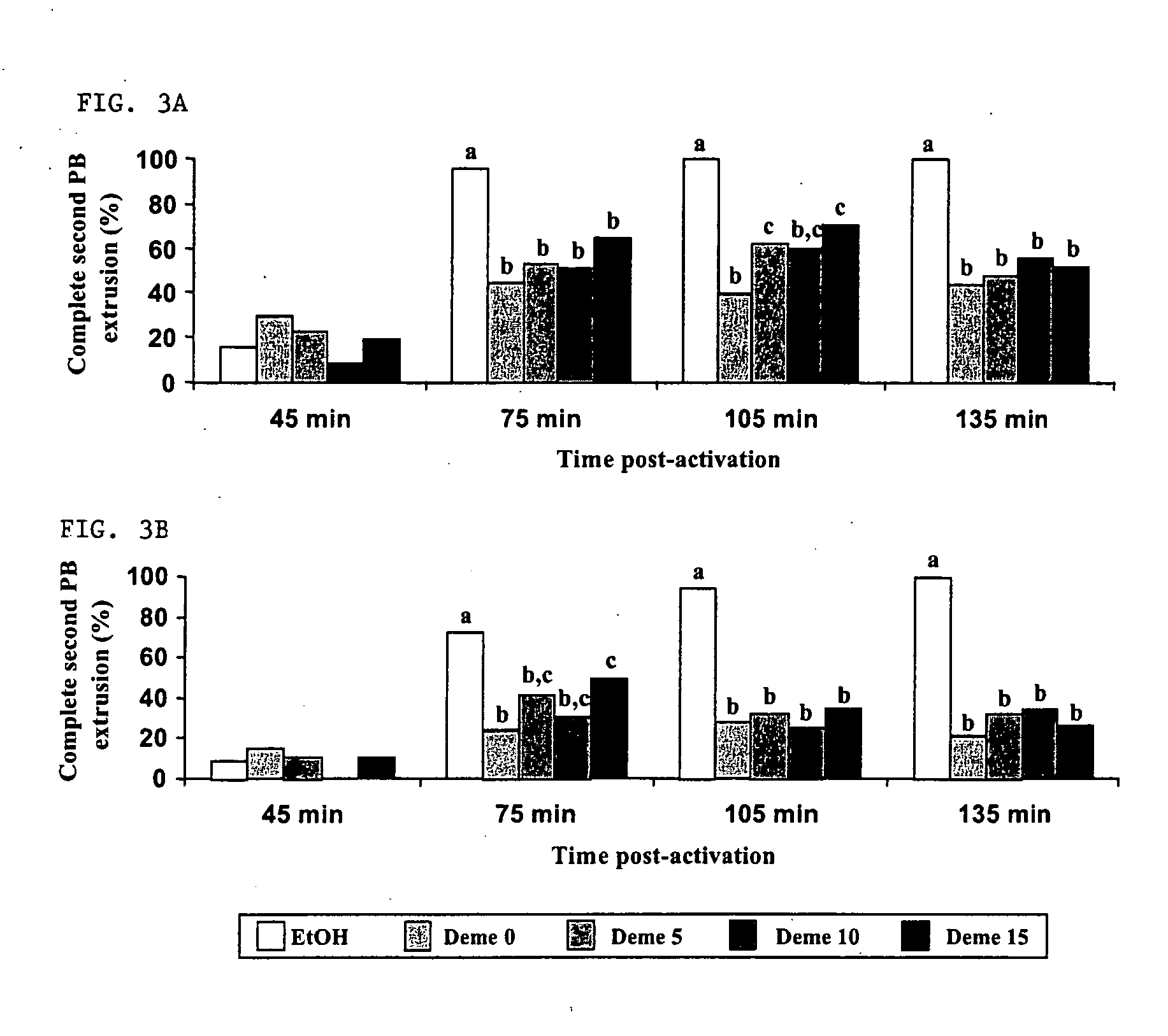
[0084] Studies were designed to further explore the use of pharmacological agents to create developmentally competent enucleated mouse oocytes for animal cloning by somatic cell nuclear transfer. Metaphase-II oocytes from CF-1 and B6D2F1 strains were activated with ethanol and subsequently exposed to demecolcine at various times post-activation. Chromosome segregation, spindle dynamics and polar body (PB) extrusion were monitored by fluorescence microscopy using DNA, microtubule and microfilament selective probes. Exposure to demecolcine did not affect rates of oocyte activation induced by ethanol but did disrupt the coordination of cytokinesis and karyokinesis, suppressing the extent and completion of spindle rotation and second PB extrusion in a strain-dependent manner. Moreover, strain and treatme...

example 3

Activated Bovine Cytoplasts Produced by Induced Enucleation Support Development of Nuclear Transfer Embryos In Vitro

[0115] Poor efficiency of somatic cell NT has been associated with the preparation of developmentally competent enucleated cytoplasts. Induced enucleation (IE) of mouse oocytes has been shown to support enhanced term development of cloned mice. This study characterized the kinetics and phenotypic progression of bovine oocytes subjected to IE, and evaluated their developmental competence to support NT embryo development in vitro. In vitro matured (26 h) oocytes were denuded, activated (5 pM ionomycin, 5 min, then 10 μg / mL cycloheximide, 5 h) and cultured for up to 5 h post-activation (pa). Oocyte enucleation was induced by demecolcine (0.4 μg / ml, DM) exposure at 30, 60, 90 and 120 min post activation for various time periods (1 to 4.5 h). Activation rates and meiotic progression of control and DM treated oocytes (n=31-49 / gp) was evaluated at 5 hpa by immunofluorescence...


PUM

PropertyMeasurementUnit
time-aaaaaaaaaa
time-aaaaaaaaaa
time-aaaaaaaaaa

Abstract

A nuclear transfer embryo is formed by destabilizing microtubules of an oocyte, whereby essentially all endogenous chromatin collects at a second polar body during meiosis of an oocyte. The oocyte is fused with the nucleus of a donor somatic cell of the same species of said oocyte prior to cessation of extrusion of the second polar body from the oocyte, thereby forming the nuclear transfer embryo. In one embodiment, the nuclear transfer embryo is employed to impregnate an animal, such as a mammal. In another embodiment, the donor nucleus is transgenic.

Description

RELATED APPLICATION [0001] This application is a continuation of International Application No. PCT / US03 / 23464, which designated the United States and was filed Jul. 29, 2003, published in English, which claims priority to and is a continuation-in-part of U.S. application Ser. No. 10 / 208,653, filed Jul. 29, 2002, the entire teachings of which are incorporated herein by reference.GOVERNMENT SUPPORT [0002] This invention was supported, in whole or in part, by Grant No. 2001-35205-09966 from the United States Department of Agriculture. The Government has certain rights in the invention.BACKGROUND OF THE INVENTION [0003] Nuclear transfer methods have been developed and used successfully to produce cloned sheep, cattle, mice, goats and pigs. Two cell components are combined to produce a cloned embryo; the donor nuclear genome (karyoplast) that is the target for clonal replication, and the enucleated oocyte (cytoplast) whose cytoplasmic constituency is sufficiently competent to facilitate ...

Claims


Application Information

Patent Timeline
Patent Type & Authority Applications(United States)
IPC IPC(8): A01K67/027C12N15/873C12N15/877
CPCA01K2227/101C12N15/8771C12N15/873
Inventor OVERSTROM, ERIC W.RUSSELL, DANIELA FISCHER
Owner TRUSTEES OF TUFTS COLLEGE
Why Eureka
Try Eureka