
Compound binding to leukocytes and medicinal composition containing the compound in labeled state as the active ingredient

a technology of compound binding and leukocytes, which is applied in the direction of drug compositions, peptides, immunological disorders, etc., can solve the problems of insufficient radiation energy of gamma rays from sup>67/sup>ga, inability to obtain good photographic images by a common gamma camera, and requiring waiting time of about 72 hours

Inactive Publication Date: 2006-03-16
NIHON MEDI PHYSICS CO LTD
View PDF3 Cites 1 Cited by
  • Summary
  • Abstract
  • Description
  • Claims
  • Application Information

AI Technical Summary

Benefits of technology

[0038] In consideration of the circumstances of prior art, an object of the present invention is to provide a compound having binding properties specific to leukocytes, i.e., neutrophils, monocytes and lymphocytes both in vivo and in vitro and capable to be labeled with a radioactive metal or a paramagnetic metal, and a medicinal composition containing said compound in labeled state as the active ingredient which is useful for SPECT image diagnosis, PET image diagnosis, MRI image diagnosis, radioactive treatment and so on, wherein imaging is performed in a site with vigorous leukocyte infiltration accompanied by an immune reaction in an individual.

Problems solved by technology

However, since the regulation of the reaction in an individual is imperfect with respect to the purpose of maintaining its biosis, the reaction may sometime cause crucial negative effects.
However, in the case of autoimmune diseases, there is no specific cause factor, or the factor has not been identified.
However, this agent has no specificity for infection site or inflammation site in addition, the radiation energy of gamma ray from 67Ga is insufficient and not suitable for obtaining a good photographic image by a common gamma camera.
Further, it requires waiting time for about 72 hours from injection of the agent to complete imaging.
However, when 111In-leukocyte is utilized, preparation of a radioactive labeled compound requires steps of aseptic withdrawal of autologous blood, aseptic isolation of leukocytes from the blood, aseptic labeling of leukocytes, and back injection of leukocytes labeled with radioactive nuclide to the patient, and takes a considerable time for more than 2 hours.
Further, radioactivity of 200 μCi per 1×10⁷ cells of leukocytes which is normally used for imaging studies is hazardous for leukocytes when In-111 is employed because of its high radioactive energy.
Therefore, it is difficult to monitor dynamic states of lymphocytes and monocytes using In-111 labeled leukocytes.
Furthermore, from the viewpoint of the safe radiation dosage, an administration quantity of radioactivity is limited, in many cases resulting in deterioration of pictorial image quality.
However, since the labeling stability of Tc-99m is not good enough, accumulation in a non-target metabolic organ becomes a problem accordingly.
However, 111In-HIG has similar drawback with respect to the waiting period of 24 to 48 hours from administration for obtaining optimal pictorial image.
However, from the viewpoint that these proteins including antibodies have large molecular weights, the diapedesis of these proteins with blood components may cause problems in acute inflammation which with enhanced vascular permeability (Roitt, I. et al., Essential Immunology, 8th edn.
In addition, use of heparin has sometime to be limited because of its physiological activity.
In addition to this, there may be a risk of side effects brought by physiological function of heparin including, for example, antithrombin activity, inhibition of thromboplastin production, and inhibition of platelet aggregation, or careful treatment is required when the patient has hemorrhagic disease or a potential to become hemorrhagic or severe liver disease and kidney disease.

Method used


Image

  • Compound binding to leukocytes and medicinal composition containing the compound in labeled state as the active ingredient
  • Compound binding to leukocytes and medicinal composition containing the compound in labeled state as the active ingredient
  • Compound binding to leukocytes and medicinal composition containing the compound in labeled state as the active ingredient

Examples


example 1

Synthesis of Peptides

[0123] Following peptides were synthesized by the solid phase peptide synthesis and used in Examples hereinbelow.

Compounds Binding to Leukocytes of the Present Invention

[0124] Peptide 3: formyl-Nle-Leu-Phe-Nle-Tyr-Lys(NH2)-ε-(-Ser-Cys-Gly-Asn); [0125] Peptide 4: formyl-Nle-Leu-Phe-Nle-Tyr-Lys(NH2)-ε-(-Ser-Cys-Asp-Asp); [0126] Peptide 5: formyl-Nle-Leu-Phe-Nle-Tyr-Lys(NH2)-c-(-Ser-Cys-Gly-Asp); [0127] Peptide 6: formyl-Nle-Leu-Phe-Nle-Tyr-Lys(NH2)-ε-(-Ser-D-Arg-Asp-Cys-Asp-Asp); [0128] Peptide 7: formyl-Nle-Leu-Phe-Nle-Tyr-Lys(NH2)-ε-(-Ser-cyclam tetracarboxylic acid); [0129] Peptide 8: formyl-Nle-Leu-Phe-Lys(NH2)-ε-(-Ser-D-Ser-Asn-D-Arg-Cys-Asp-Asp); [0130] Peptide 9: formyl-Nle-Leu-Phe-Nle-Tyr-Lys(NH2)-ε-(-Ser-D-Arg-DTPA); [0131] Peptide 13: formyl-Nle-Leu-Phe-Nle-Tyr-Lys(NH2)-ε-(-Ser-Cyclam butyric acid); [0132] Peptide 14: formyl-Nle-Leu-Phe-Nle-Tyr-Lys(NH2)-ε-(-Ser-D-Arg-Asp-cyclam butyric acid); [0133] Peptide 15: formyl-Nle-Leu-Phe-Nle-Tyr-Lys(NH2)-ε-...

example 2

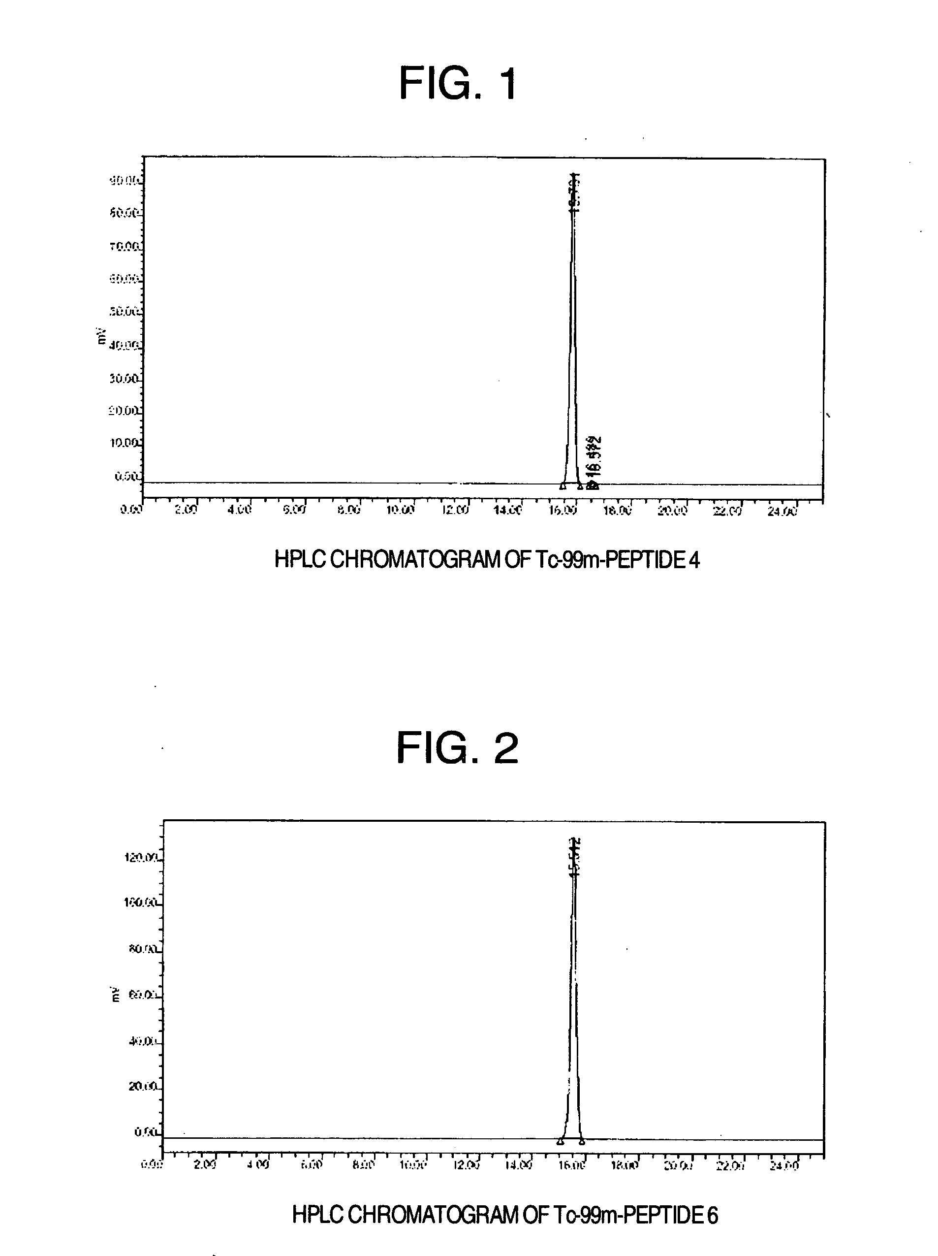
Tc-99m Labeling of Peptide 1, Peptide 2, Peptide 3, Peptide 4, Peptide 5, Peptide 6, Peptide 7, Peptide 8, Peptide 9, Peptide 10, Peptide 13, Peptide 14, Peptide 15, Peptide 16, Peptide 17 and Peptide 18

(1) Method

[0166] A solution of Tc-99m-sodium pertechnetate (hereinafter designated as 99mTcO4−), 1.1-3.0 GBq, was added into a vial containing a mixture of glucoheptonic acid 40.3 μmol / 300 μl and stannous chloride solution 130 nmol / 50 μl to make the total volume 1.35 ml. The mixture was reacted at room temperature for 30 minutes with stirring and occasional tumbling. A part thereof was collected and the labeling rate of Tc-99m of Tc-99m-glucoheptonic acid was confirmed to be 95% or more.

[0167] Each of sixteen peptides obtained in Example 1 was dissolved in dimethylformamide (DMF) and a concentration thereof in each solution was adjusted to 0.25-12.5 nmol / 200 μl with ultra pure water, 10 mM phosphate buffer containing 0.9% NaCl, pH 7.4, (hereinafter designated as PBS) or 10 mM ca...

example 3

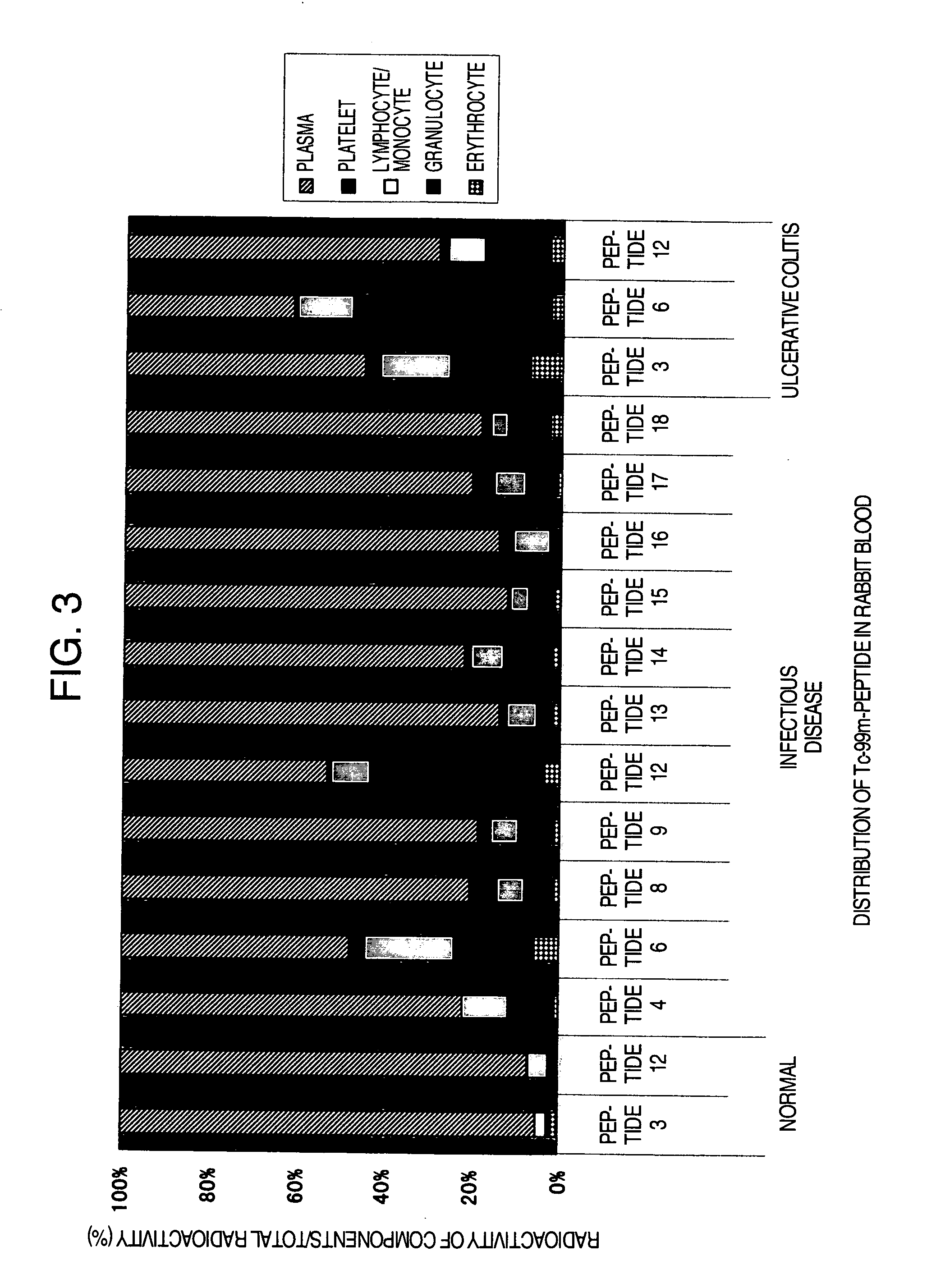
Distribution in Rabbit Blood

[0169] (1) The peptides which were labeled with Tc-99m in Example 2 (Peptide 3, peptide 4, peptide 6, peptide 8, peptide 9, peptide 12, peptide 13, peptide 14, peptide 15, peptide 16, peptide 17 and peptide 18) were purified by separating into unlabeled peptides and labeled peptides using a reversed phase HPLC under the same conditions of HPLC as in Example 2. Gradient elution was performed under the condition of 20% 50% (0.1% TFA acetonitrile / 0.1% TFA water): 0→20 minutes. Subsequently, Percoll density-gradient solution was prepared. To an undiluted Percoll solution (Pharmacia Biotech Inc.) (specific gravity 1.130 g / ml) 90 ml, 1.5 M NaCl 10 ml was added to prepare an isotonic solution equal to the physiological saline. This solution was diluted by adding physiological saline to prepare 1.096, 1.077 and 1.063 g / ml of Percoll solutions. The thus prepared 1.096, 1.077 and 1.063 g / ml of Percoll solutions, each 1 ml, were layered over in a 15 ml tube. It wa...


PUM

PropertyMeasurementUnit
Fractionaaaaaaaaaa
Fractionaaaaaaaaaa
Fractionaaaaaaaaaa

Abstract

A compound binding to leukocytes, which comprises Met-Leu-Phe or Nle-Leu-Phe serving as the leukocyte-binding site of a formyl peptide receptor (FPR), a binding part comprising Ser or Thr elevating the binding ratios to monocytes and lymphocytes in all leukocytes, a group which can be labeled with a radioactive metal or a paramagnetic metal, and a spacer binding them shows binding properties specific to all leukocytes, i.e., neutrophils, monocytes and lymphocytes both in vivo and in vitro and can be labeled with a radioactive metal or a paramagnetic metal. Owing to these characteristics, this compound is highly useful in SPECT image diagnosis, PET image diagnosis, MRI image diagnosis and so on wherein imaging is performed in a site with vigorous leukocyte infiltration accompanied by an immune reaction in an individual.

Description

TECHNICAL FIELD [0001] The present invention relates to a compound binding to leukocytes and a medicinal composition containing the compound in labeled state as the active ingredient which can be used for targeting abnormal site as well as for understanding precisely a state of disease activity when a disease of an individual associated with immune reaction is to be diagnosed and / or treated. More in detail, the present invention relates to a novel compound having binding properties specific to leukocytes both in vivo and in vitro and can be labeled with a radioactive metal or a paramagnetic metal, which is useful for pathological imaging of a seat of disease including infectious diseases, inflammation, tumor and atheroscrelosis in the body of mammals. Also, the present invention relates to a medicinal composition containing said compound in labeled state as the active ingredient which is useful for radioisotope diagnosis, SPECT image diagnosis, PET image diagnosis, MRI image diagnos...

Claims


Application Information

Patent Timeline
IPC IPC(8): A61K51/00A61K38/12A61K38/00A61K38/08A61K49/14A61K51/08A61P37/00C07K7/02C07K7/06
CPCA61K38/08A61K49/14C07K7/06C07K7/02A61K51/088A61P1/00A61P1/04A61P17/00A61P19/02A61P19/04A61P29/00A61P37/00A61P37/02Y02A50/30A61K38/00A61K49/00
Inventor SEKI, IKUYAKAWAGUCHI, TAKAYOSHISHIRAKAMI, YOSHIFUMI
Owner NIHON MEDI PHYSICS CO LTD
Learn More