Target activated microtransfer

a microtransfer and target technology, applied in the field of biomolecule analysis, can solve the problems of manual labor, not highly susceptible to automation for high-throughput analysis of multiple specimens, and achieve the effects of improving the efficiency of transfer microdissection, improving the collection of cells, and high-thought-out analysis methods

Active Publication Date: 2006-06-22
HEALTH & HUMAN SERVICES GOVERNMENT OF THE UNITED STATES OF AMERICA AS REPRESENTED BY THE SERCRETARY OF THE +1
View PDF55 Cites 23 Cited by
  • Summary
  • Abstract
  • Description
  • Claims
  • Application Information

AI Technical Summary

Benefits of technology

[0009] The present disclosure describes methods, systems and devices for analyzing a biological sample, such as a cellular specimen. In one aspect of the method, a proximal activating event comes from within the biological sample for focally altering an adjacent transfer surface for selective removal of a target from within the sample. In situ activation from within the sample avoids the necessity of selecting each target and then altering the transfer surface by application of external energy, as in laser capture mic...

Problems solved by technology

Original microdissection techniques involved painstaking (and sometimes clumsy) manual dissection using needles or other micro-manipulation devices to isolate individual cells based on visible, histological characteristics.
These methods share the same inherent disadvantages of LCM, in that individual cells must be visu...

Method used


Image

  • Target activated microtransfer
  • Target activated microtransfer
  • Target activated microtransfer

Examples


example 1

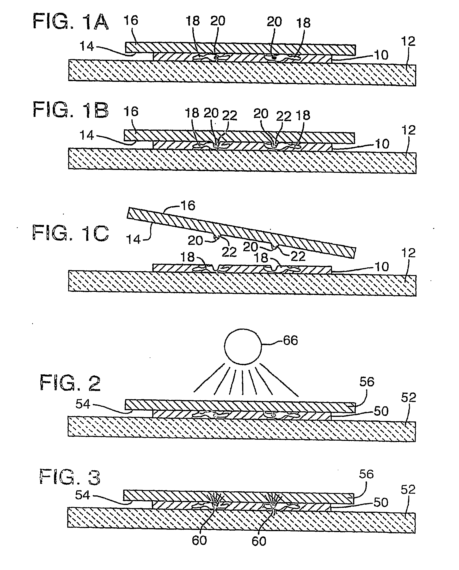
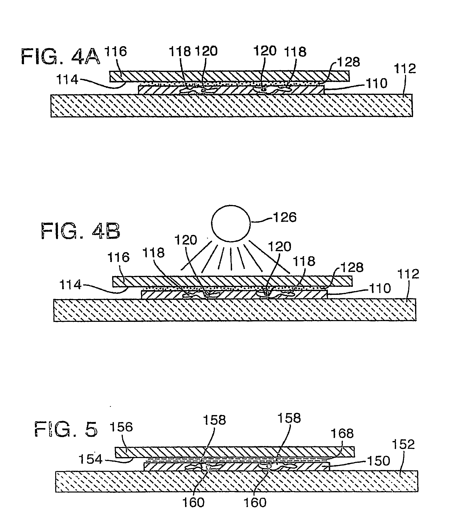
[0102] As noted above, target activated microtransfer is capable of procuring pure populations of cells that are selected according to their expression pattern. These expression patterns may be determined in a complex tissue section using, for example, immunohistochemistry (antibody detetection), or nucleic acid hybridization (with DNA or RNA binding probes). The method disclosed in this example uses standard immunohistochemistry to stain the cells of interest. Once the cells are stained, the tissue section is covered by a clear Ethylene Vinyl Acetate (EVA) film and a laser is then run across the entire tissue section. The areas that are stained absorb the energy locally and melt the EVA film. The film is then lifted off the tissue section, thereby isolating the cells of interest from the remaining tissue section.

[0103] Conventional laser-based microdissection depends on the morphological recognition of the desired cells to target them, and can be quite labor intense, particularly ...

example 2

Automated Scanning

[0119] Target activated microtransfer was performed using a 400 mW fiber optic LD diode source in the continuous mode to scan the bottom of an immunostained prostate tissue section. The fiber of the light source was in contact with the lower glass surface of the slide, while the tissue was mounted on the top glass surface. A large sheet of clear Elvax510 EVA on top of the tissue served as the transfer film. The fiber provided a 200 μm spot on the tissue section, and was scanned at ˜3 cm / sec across the lower surface of the slide to achieve fusion and specific microtransfer of target.

[0120] Microtransfer can be automated in this fashion, for example by using a programmable stage to move the tissue or a laser scanner relative to the slide. This programmed or predetermined movement sweeps the laser beam across the tissue, allowing the entire slide to be irradiated in ˜1 minute.

[0121] Many other laser sources could be used for this purpose, such as a 1 W argon laser....

example 3

Examples of Tested Targets

[0122] A variety of different primary antibodies were tested in the disclosed microdissection / microtransfer method to demonstrate the general applicability of the method to a variety of different targets. Antibodies tested include: Cyokeratin AE1 / AE3; LP34 (Cytokeratin, Basal Cells; CD34; CD3; PSA; Desmin; E-cadherin; S-100; GAPDH; CD45RO; Histone; Vimetin; and Actin.

[0123] All of these antibodies provided specific activation and adherence to the transfer layer, with no melting observed in unstained areas.


PUM

PropertyMeasurementUnit
Chemical shiftaaaaaaaaaa
Temperatureaaaaaaaaaa

Abstract

A method of removing a target from a biological sample which involves placing a transfer surface in contact with the biological sample, and then focally altering the transfer surface to allow selective separation of the target from the biological sample. In disclosed embodiments, the target is a cell or cellular component of a tissue section and the transfer surface is a film that can be focally altered to adhere the target to the transfer surface. Subsequent separation of the film from the tissue section selectively removes the adhered target from the tissue section. The transfer surface is activated from within the target to adhere the target to the transfer surface, for example by heating the target to adhere it to a thermoplastic transfer surface. Such in situ activation can be achieved by exposing the biological sample to an immunoreagent that specifically binds to the target (or a component of the target). The immunoreagent can alter the transfer surface directly (for example with a heat generating enzyme carried by the immunoreagent), or indirectly (for example by changing a characteristic of the target). In some embodiments, the immunoreagent deposits a precipitate in the target that increases its light absorption relative to surrounding tissue, such that the biological specimen can be exposed to light to selectively heat the target. Alternatively, the immunoreagent is an immunofluorescent agent that carries a fluorophore that absorbs light and emits heat.

Description

CROSS REFERENCE TO RELATED APPLICATION [0001] This application claims the benefit of U.S. Provisional Application No. 60 / 442,399, filed Jan. 24, 2003, which is incorporated herein in its entirety.FIELD OF THE DISCLOSURE [0002] The present disclosure is related to methods of analysis of biomolecules, particularly methods that involve targeting components of biological samples for subsequent separation of components of interest from their surroundings. BACKGROUND [0003] A variety of techniques have been used to microdissect specific cells or cell populations from a histological sample under direct microscopic visualization. Original microdissection techniques involved painstaking (and sometimes clumsy) manual dissection using needles or other micro-manipulation devices to isolate individual cells based on visible, histological characteristics. [0004] More recent techniques have been developed to more efficiently separate biological components, such as particular subsets of cells, from...

Claims


Application Information

Patent Timeline
IPC IPC(8): G01N33/53G01N33/537G01N33/543G01N33/569
CPCG01N33/543Y10T428/2839Y10T428/2813G01N33/56966
Inventor EMMERT-BUCK, MICHAEL R.TANGREA, MICHAEL ANTHONYBONNER, ROBERT F.CHUAQUI, RODRIGO F.
Owner HEALTH & HUMAN SERVICES GOVERNMENT OF THE UNITED STATES OF AMERICA AS REPRESENTED BY THE SERCRETARY OF THE
Why Eureka
Try Eureka