In-gel fluorescent protein staining technique

a protein staining and gel technology, applied in the direction of fluid pressure measurement, liquid/fluent solid measurement, peptides, etc., can solve the problems of inconvenient post-electrophoresis gel staining and destaining, loss of protein band signal, time and money, etc., to reduce the post-electrophoretic process time and improve yield

Inactive Publication Date: 2006-07-06
KITZLER JEFFREY +2
View PDF0 Cites 8 Cited by
  • Summary
  • Abstract
  • Description
  • Claims
  • Application Information

AI Technical Summary

Benefits of technology

[0021] Since staining occurs during the run, post-electrophoretic manipulations of the gel are minimized. As compared to the standard Coomassie staining technique, the system of the invention reduces post-electrophoretic process time from 4-8 hours to 30 minutes. When compared to existing fluorescent protein stains, the inventive system is generally faster and substantially cheaper.
[0022] The non-denaturing aspect of the stain allows proteins to be recovered from the gel with substantially improved yields relative to other procedures currently available. For example, the user can rec...

Problems solved by technology

However, the separated proteins are not generally visible to the naked eye.
The difference in visibility often depends on how well the gel is destained but excessive destaining can result in loss of the protein band signal.
Moreover, the post-electrophoresis manipulation of the gels for staining and destaining is inconvenient and costs time and money.
In addition to the foregoing, the staining and dest...

Method used


Image

  • In-gel fluorescent protein staining technique
  • In-gel fluorescent protein staining technique

Examples


example 1

[0051] A gel containing the protein specimen(s) is mounted in a standard electrophoresis apparatus. The lower buffer chamber is filled with the 1× Tris-Glycine SDS (0.192 Glycine, 0.025 M Tris, and 0.1% SDS). The upper buffer chamber is filled with the running buffer, which in this case is the detection reagent containing Nile red described hereinabove.

[0052] The gel is run under standard voltage and temperature conditions, typically at 175 Volts for 1 hour.

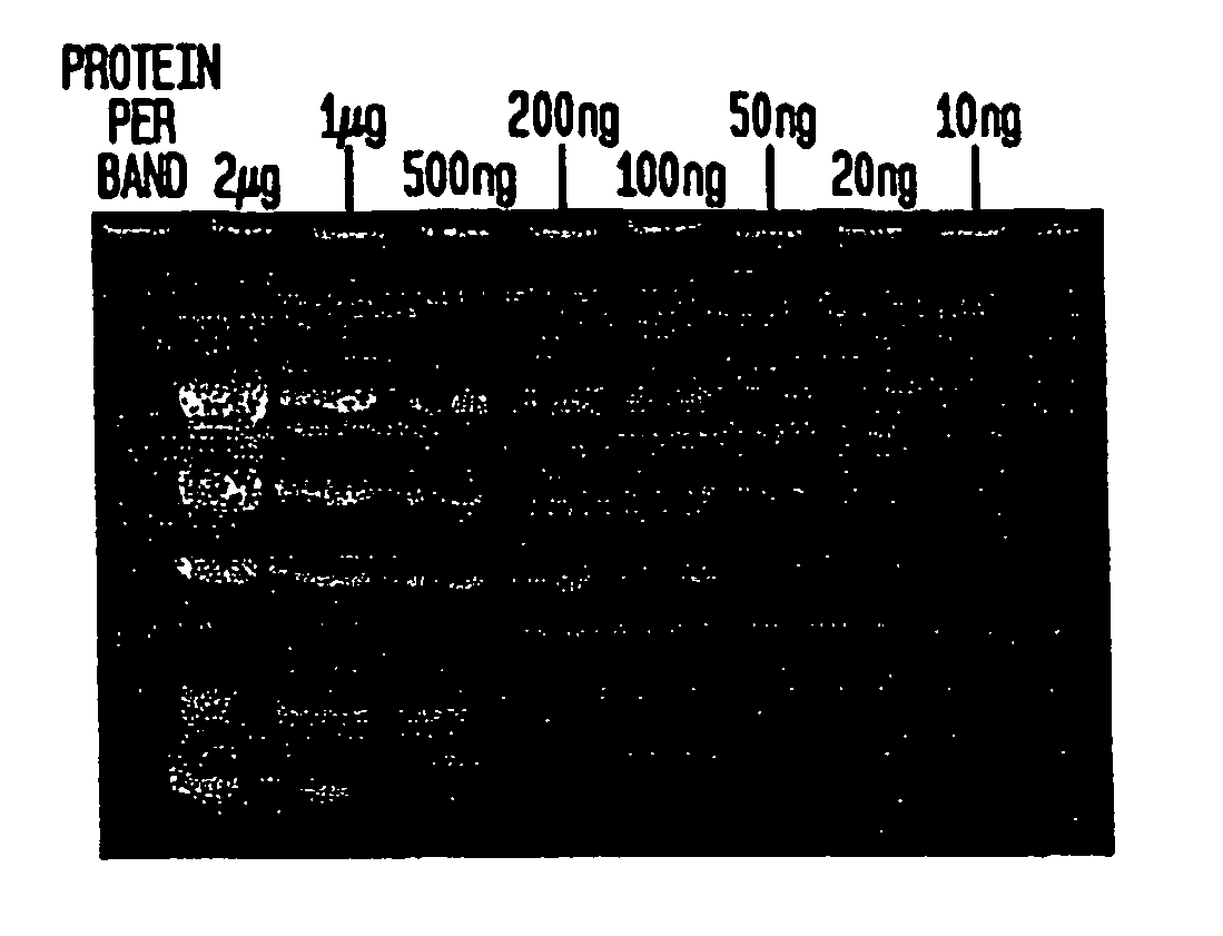
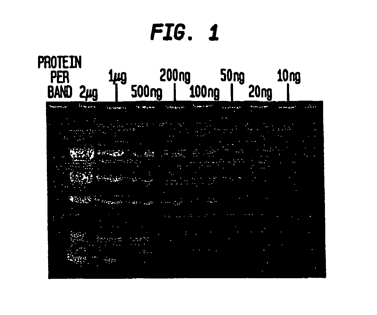
[0053] When the gel is removed from the electrophoresis apparatus, the separated protein bands can be visualized immediately using a transilluminator under ultraviolet illumination (302 nm) for the detection of bands containing more than 300 ng of protein. Destaining increases sensitivity. In the preferred method of destaining, the gel is washed, or immersed, in 50 ml deionized water for 15 minutes followed by a second wash in 50 ml deionized water for 15 minutes.

[0054] The gel is observed on a transilluminator (302 nm). Prote...

example 2

[0057] In an alternative embodiment, the destaining solution is an aqueous solution of 0.1 M potassium chloride (KCl). The KCl solution is used in lieu of deionized water in the destaining protocol described hereinbove.

example 3

[0058] In another practical embodiment of the invention, the detection reagent contains a Phosphine dye.

[0059] A gel containing the protein specimen(s) is mounted in a standard electrophoresis apparatus. The lower buffer chamber is filled with the 1× Tris-Glycine SDS buffer. The upper buffer chamber is filled with a detection reagent made from the Phosphine Concentrate described above (10 mg Phosphine dye in 1 ml water) added to 150 ml 1× Tris-Glycine SDS buffer.

[0060] The gel is run under standard voltage and temperature conditions, typically at 150 Volts for 1 hour.

[0061] The gel is destained in 0.0M KCl for 10 minutes and photographed using a UV transilluminator with a green filter.

[0062] The detection limit for the technique of Example 3 is between 50 ng-100 ng per protein band.


PUM

PropertyMeasurementUnit
Fractionaaaaaaaaaa
Fractionaaaaaaaaaa
Fractionaaaaaaaaaa

Abstract

An “in-gel” staining technique for detecting and/or separating proteins during electrophoresis, illustratively as a modification of the standard Laemmli procedure. A fluorescent dye, such as Nile red or Phosphine, is included in the running buffer (mobile phase) which may be a standard Laemmli Tris-Glycine SDS buffer that has been modified to reduce the concentration of detergent (SDS) to less than the typical concentration (0.10% v/v). The fluorescent dye stains proteins during electrophoretic separation. The post-electrophoretic operations are, therefor, reduced and the separated, stained fractions are recoverable for further processing, purifying or analysis.

Description

CROSS-REFERENCE TO RELATED APPLICATIONS [0001] This application claims the benefit under 35 U.S.C. § 119(e) of U.S. Ser. No. 60 / 421,021 filed Oct. 23, 2002. The disclosures of this application is incorporated herein by reference.BACKGROUND OF THE INVENTION [0002] 1. Field of the Invention [0003] This invention relates generally to a system for detecting proteins by electrophoresis, and more particularly to a system for the staining of proteins with a fluorescent dye during electrophoretic separations. [0004] 2. Description of the Related Art [0005] One of the most widely used methods for the separation of proteins is gel electrophoresis. A sample of a protein on an inert support is subjected to an electric field that causes the proteins to migrate in accordance with molecular weight. Typically, the supports are made of polymers, such as polyacrylamide, which is a copolymer of acrylamide and bisacrylamide, or agarose, a polymer of glucose units. In the most common method of electroph...

Claims


Application Information

Patent Timeline
IPC IPC(8): B01D57/02B01D59/42G01N27/447
CPCC07K1/26G01N27/44726
Inventor KITZLER, JEFFREYWERNER, DAVIDIBRAHIM, ABDUL
Owner KITZLER JEFFREY
Why Eureka
Try Eureka