
High throughput use-dependent assay based on stimulation of cells on a silicon surface

Inactive Publication Date: 2007-03-01
NEUROSILICON 1145990 ALBERTA
View PDF26 Cites 22 Cited by
  • Summary
  • Abstract
  • Description
  • Claims
  • Application Information

AI Technical Summary

Benefits of technology

[0021] The invention also provides a method for measuring the effect of a test compound on cellular activity, the method comprising: providing a silicon wafer having a surface suitable for cell growth; culturing at least one cell on the surface; contacting the at least one cell with a test compound; repetitive

Problems solved by technology

Such multiple uses of a channel are said to wear the channel down.
Although effective, this approach allows the analysis of only one region of one cell at any given time and requires significant skilled human labor.
However, the utility of the transistor array is constrained by the fixed spatial position of the transistor elements; the positions of individual neurons in a neuronal network cannot be guaranteed to align perfectly with the transistor elements and thus the individual neurons cannot always be selectively stimulated.
These techniques, although offering the possibility of repetitive stimulation of cells, suffer from the drawback that the cells must be pre-incubated with a special compound that releases the neurotransmitter when excited by a laser.
These techniques are therefore invasive.
Uncaging complicates interpreting the results of these measurements because the neurotransmitter diffuses away from its normal synaptic localization.
Furthermore, there are limitations on the duration of stimulation that is possible.
However, hitherto it has only been used as a research tool for investigating structural changes that result from long-term cellular excitation, for example, in elucidating how long-term memories are established by neuronal networks.
However, to date, photoconductive stimulation of cells has not formed the basis of a screening assay.
However, hitherto these two types do not allow assaying of drugs that require repetitive stimulation of ion channel activity in order to reach their maximum effects because an underlying way of repetitively stimulating cells has not been available in the respective assay format.
The drawback of this assay in the context of ion channel activity is that it is a static test that is not based on the measurement of a drug's effects on ion channel activity per se.
In other words, even if a drug binds to the channel, the drug may not be biologically active because the cell may not have been appropriately stimulated.
This invalidates many of the findings from a radioligand binding assay.
One major drawback of this type of assay is that each cell is depolarized only once, by immersion in, e.g., KCl, and for a prolonged time, thus rendering the culture incapable of subsequent screening through subsequent stimulation because the KCl cannot be removed.
Another major drawback of this type of assay is that prolonged depolarization causes excito-toxicity in neuronal cells that arises when a cell is overstimulated or depolarized for too long, triggering apoptotic processes and causing the cell to die.
Neuronal excito-toxicity and apoptotic processes might introduce confounding factors into these types of experiments and lead to flawed results.
This technique, with the aforementioned limitations, does not allow for the screening of compounds that inhibit ion channels by a mechanism called use-dependent block.
The conventional fluorescent based and radioligand assays do not allow for repetitive cell stimulation, but rather stimulate the cell only once.
Thus, many potentially useful drugs cannot be identified by either of these conventional screening processes.
Patch clamping is also not practical for testing potential drug candidates.
In other words, the patch clamp system can stimulate cells repetitively but it cannot do this in a highly parallel and automated fashion.
Therefore, one of the limiting factors in current drug screening technology is the inability to find therapeutically active frequency-dependent ion channel inhibitors easily and quickly using automated testing systems.
Hitherto, assays using such systems are either limited to stimulating cells only once or are not able to selectively stimulate particular cells.
Consequently, although currently available robotic screening devices can test many compounds quickly and automatically they cannot stimulate cells repetitively.
Hitherto, the photoconductive stimulation method has not been adapted for high throughput screening.

Method used


Image

  • High throughput use-dependent assay based on stimulation of cells on a silicon surface
  • High throughput use-dependent assay based on stimulation of cells on a silicon surface
  • High throughput use-dependent assay based on stimulation of cells on a silicon surface

Examples


example 1

Assay Apparatus

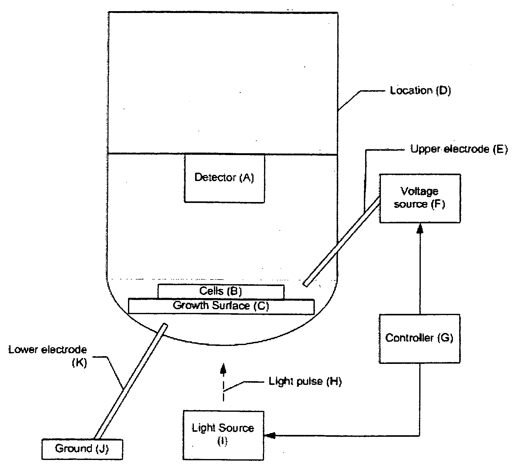
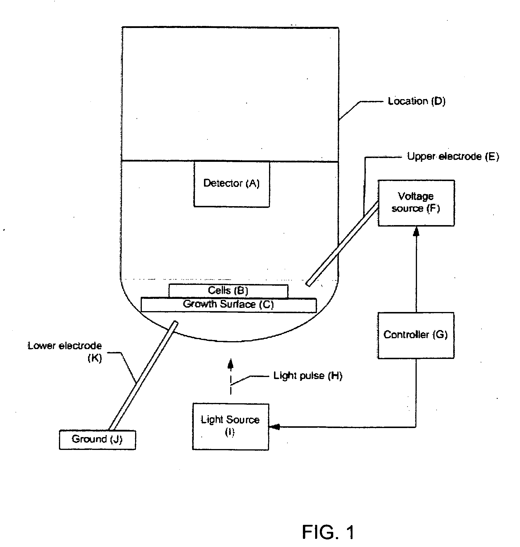
[0095] The components of a 96-well assay system, according to one embodiment of the invention, are shown in FIGS. 3-9. Although the example system contains 96 wells, the same approach could be applied to a system with a different number of wells, for example, 64, 100, 128, 256, or 512. In the representative embodiment (shown in cross-sectional view from the side in FIGS. 8A and 8B), the system is comprised of three major subassemblies: a bottom plate assembly 100 containing the light sources used for the photoconductive stimulation; an assembly having a middle plate 130 containing the wells, an array of silicon wafers 120, and upper and lower electrodes, and a carrier plate 100; and an upper plate assembly 140 containing the fluorescence excitation light source, dichroic mirror and filter assembly, and the fluorescence imagers. FIG. 6 shows an array of wells 132 disposed in middle plate 130. FIG. 5 shows a wafer assembly layer 120 having an array of individual wafers...

example 2

Photoconductive Stimulation of Human Embryo Kidney Cells

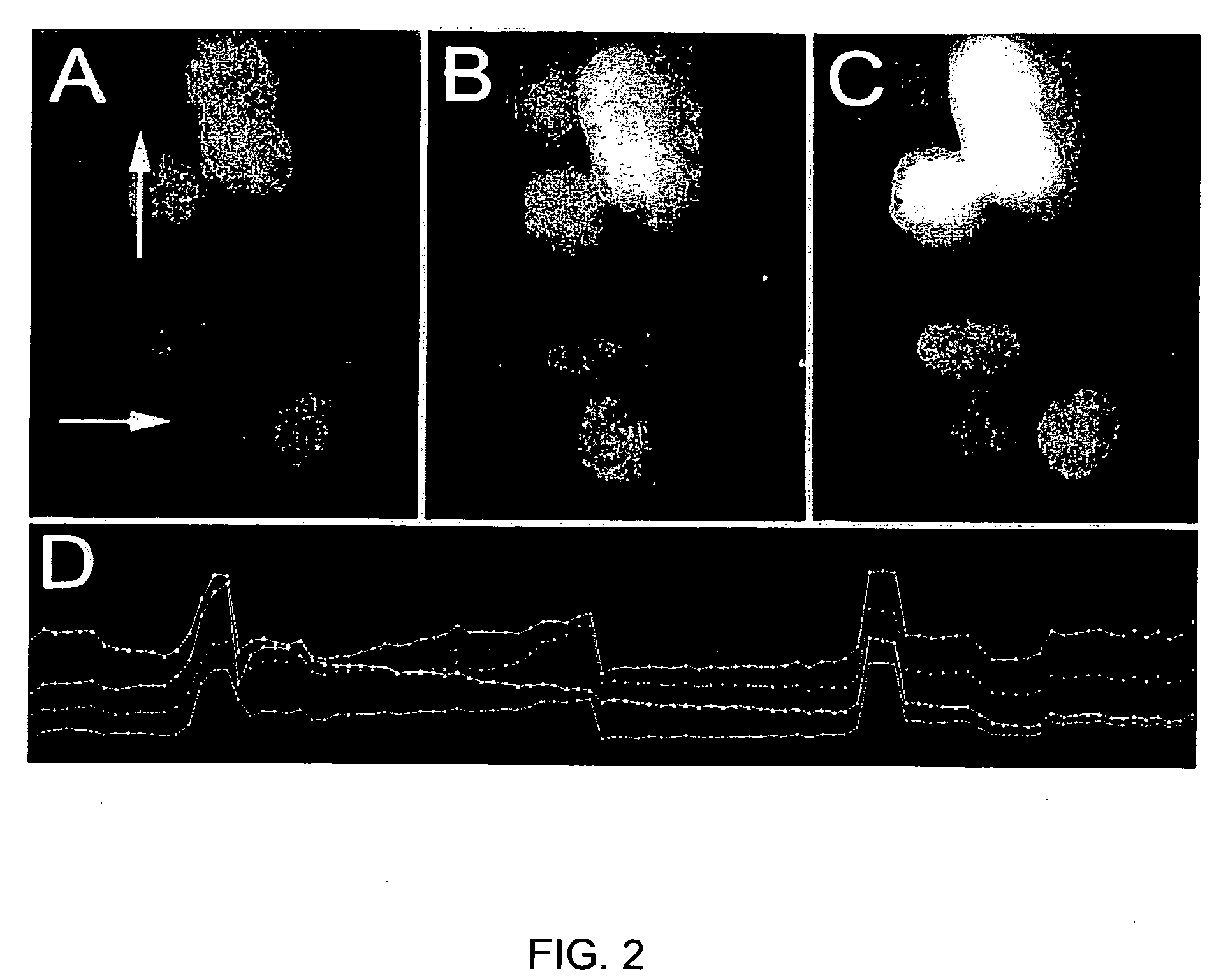
[0103] In FIG. 2, images A-C show Fluo4 calcium imaging of HEK cells which have been transfected with calcium channel subunits before (A) and after (B, C) depolarization with photoconductive stimulation. An increase in the level of calcium within the cells can be observed by the increase in fluorescence. Arrows identify cells that change. Panel (D) is a graph that demonstrates quantitative measurement of calcium indicator signal levels in a subset of cells. Depolarizations can be detected by peaks on the graph.


PUM


Abstract

The invention pertains generally to the field of drug discovery science. More specifically, the invention refers to a drug screening assay and device that is used to test the effect of specific chemical compounds as use dependent ion channel blockers. The invention includes a unique method for stimulating cells with photoconductive stimulation and reading consequent cell ion channel activity with the optical imaging of fluorescent dyes.

Description

CROSS REFERENCE TO RELATED APPLICATIONS [0001] This application claims the benefit of priority under 35 U.S.C. § 119 to U.S. provisional application, having Ser. No. 60 / 683,132 filed May 20, 2005, the disclosure of which is incorporated herein by reference in its entirety. The disclosures of U.S. provisional applications having Ser. Nos. 60 / 689,645 filed Jun. 10, 2005, 60 / 691,012, filed Jun. 15, 2005, 60 / 691,322, filed Jun. 15, 2005, and 60 / 699,829, filed Jul. 15, 2005 are incorporated herein by reference in their entirety.FIELD OF THE INVENTION [0002] This invention relates to the field of drug discovery science, and more particularly to methods and devices for screening compounds based on their effect on repetitively stimulated cells on a photoconducting silicon surface. BACKGROUND [0003] Certain physiological processes rely on cells that are excitable. A cell is excitable if it generates an action potential, which is a rapid and dramatic change in the cell's electrochemical poten...

Claims


Application Information

Patent Timeline
IPC IPC(8): C12Q1/00C12M3/00
CPCG01N21/6428G01N21/6454G01N33/5008G01N2500/10G01N33/5061G01N33/6872G01N33/5058
Inventor COLICOS, MICHAEL A.ZAMPONI, GERALD
Owner NEUROSILICON 1145990 ALBERTA
Learn More