Gene specifically expressed in postmitotic dopaminergic neuron precursor cells

a dopaminergic neuron and gene technology, applied in the field of new 65b13 gene expressed in postmitotic dopaminergic neurons, to achieve the effect of high therapeutic efficacy, convenient preparation, and efficient separation

Inactive Publication Date: 2007-05-31
EISIA R&D MANAGEMENT CO LTD
View PDF6 Cites 57 Cited by
  • Summary
  • Abstract
  • Description
  • Claims
  • Application Information

AI Technical Summary

Benefits of technology

[0014] The in situ hybridization results were further supported by immunostaining using an anti-65B 13 antibody (FIG. 13). Moreover, populations of cells expressing 65B13 could be efficiently separated by flow cytometry using an anti-65B13 antibody (FIG. 14).
[0015] According to the above results, anti-65B13 antibodies can be used to obtain pure early-stage dopaminergic neuron precursor cells, by isolating 65B13-expressing cells from ventral midbrain region or cell cultures that contain in vitro-differentiated dopaminergic neurons. Cells obtained in this manner contain only postmitotic precursor cells, and since only the cell type of interest is isolated, these cells are extremely safe even when used for transplant therapy. Since the earliest possible precursor cells are used, high therapeutic efficacy can be expected in terms of their survival rate, network formation ability, and such. Further, in the cases where the best therapeutic effects cannot be achieved by these early precursor cells obtained immediately after cell cycle exit, and where the use of matured cells is required, early precursor cells obtained by this method can simply be cultured in vitro to mature into a suitable stage of differentiation. Thus, materials that are in a differentiation stage suitable for the target transplant therapy can be easily prepared (FIG. 12).

Problems solved by technology

One of the major problems in Parkinson's disease (PD) transplant therapy at the moment is that in vitro differentiated dopaminergic neuron precursor cells and midbrain ventral zone of aborted fetuses are both mixtures of myriad types of cells.
Further, in the cases where the best therapeutic effects cannot be achieved by these early precursor cells obtained immediately after cell cycle exit, and where the use of matured cells is required, early precursor cells obtained by this method can simply be cultured in vitro to mature into a suitable stage of differentiation.

Method used


Image

  • Gene specifically expressed in postmitotic dopaminergic neuron precursor cells
  • Gene specifically expressed in postmitotic dopaminergic neuron precursor cells
  • Gene specifically expressed in postmitotic dopaminergic neuron precursor cells

Examples


example 1

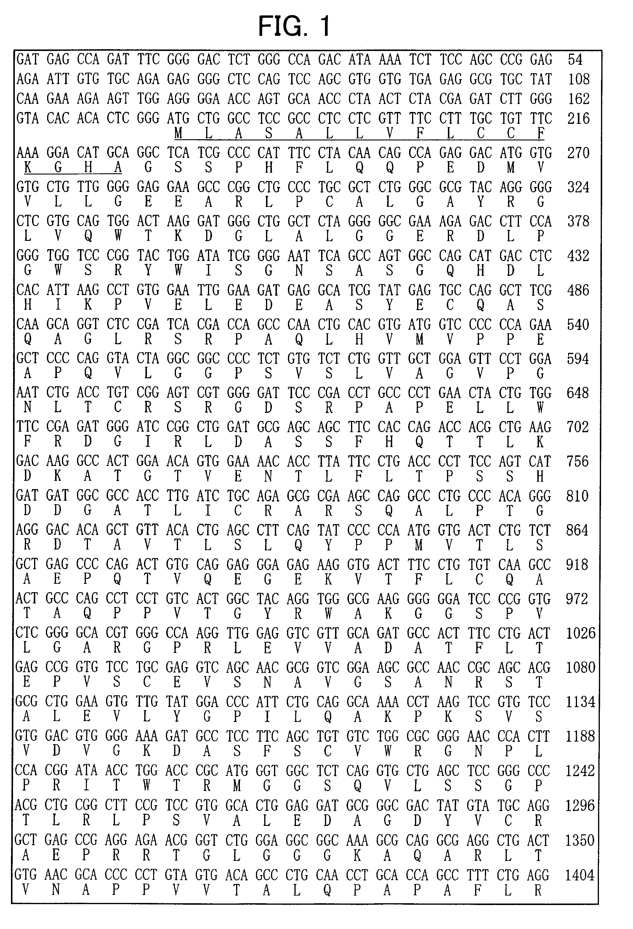
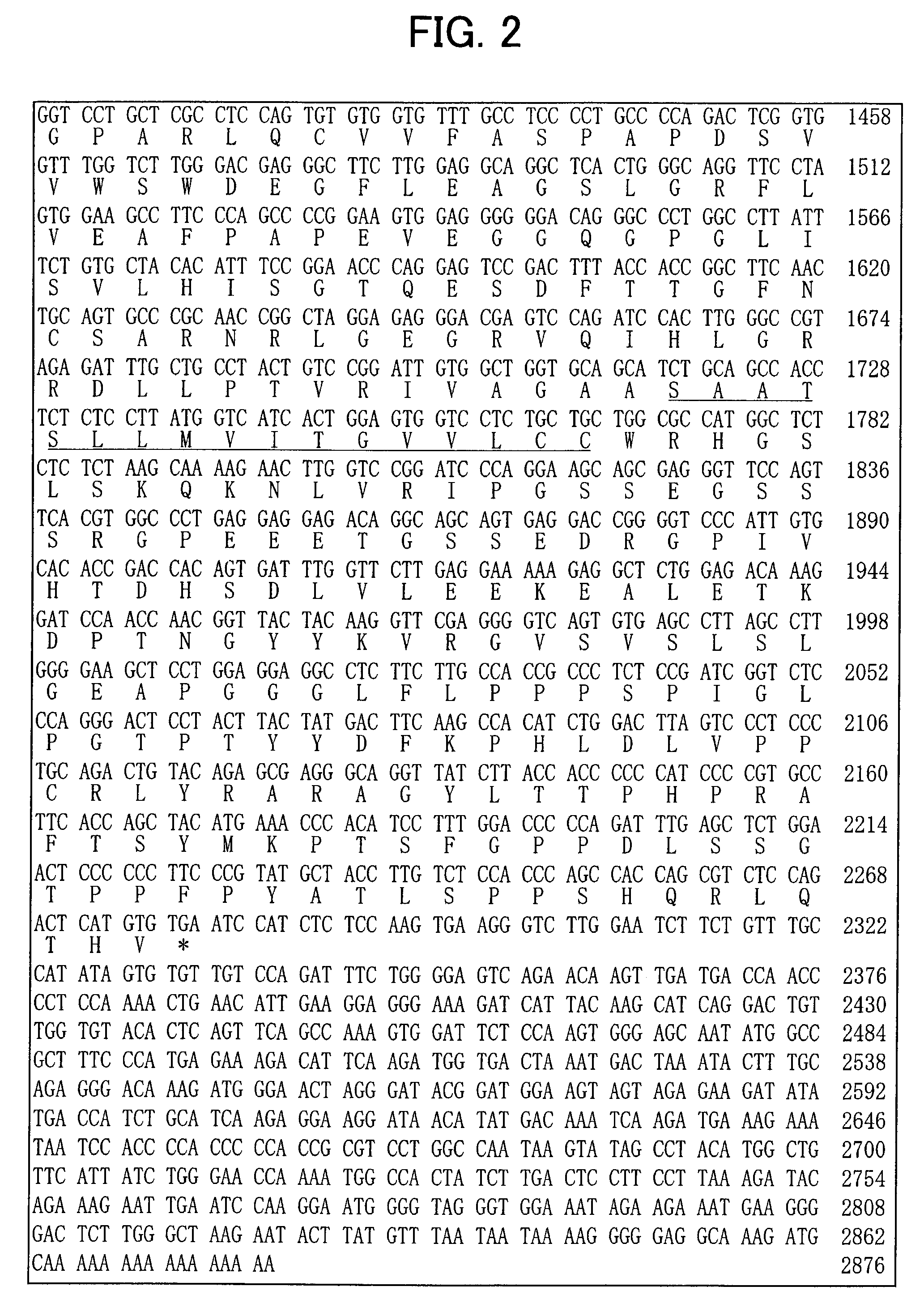
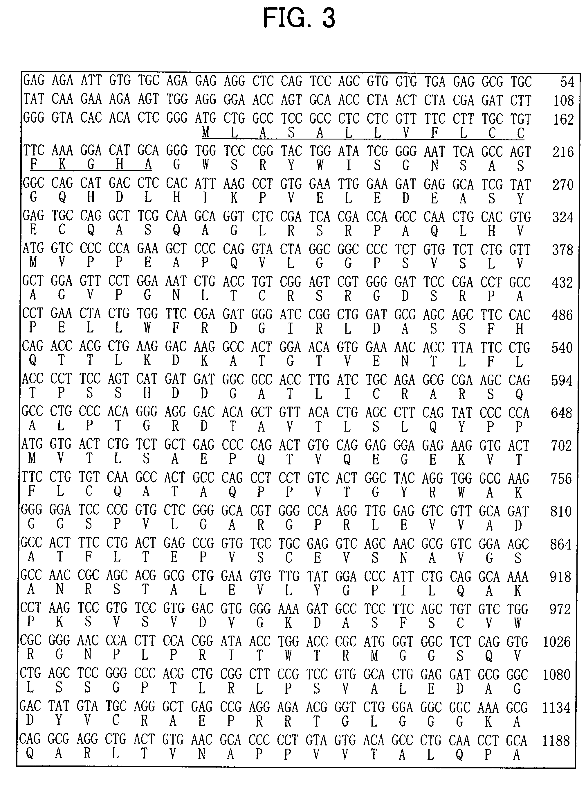
Isolation and Sequence Analysis of a Gene Specific for Dopaminergic Neuron Precursor Cells

[0123] To isolate a gene specific to dopaminergic neuron precursor cells, genes with differences in expression were amplified by the subtraction (N-RDA) method using RNA from ventral and dorsal midbrain of E12.5 mice, and sequences of the resulting genes were analyzed.

1. N-RDA Method

1-1. Adapter Preparation

[0124] The following oligonucleotides were annealed to each other, and prepared at 100 μM. (ad2: ad2S+ad2A, ad3: ad3S+ad3A, ad4: ad4S+ad4A, ad5: ad5S+ad5A, ad13: ad13S+ad13A)

ad2S:cagctccacaacctacatcattccgt(SEQ ID NO:11)ad2A:acggaatgatgt(SEQ ID NO:12)ad3S:gtccatcttctctctgagactctggt(SEQ ID NO:13)ad3A:accagagtctca(SEQ ID NO:14)ad4S:ctgatgggtgtcttctgtgagtgtgt(SEQ ID NO:15)ad4A:acacacteacag(SEQ ID NO:16)ad5S:ccagcatcgagaatcagtgtgacagt(SEQ ID NO:17)ad5A:actgtcacactg(SEQ ID NO:18)ad13S:gtcgatgaacttcgactgtcgatcgt(SEQ ID NO:19)ad13A:acgatcgacagt.(SEQ ID NO:20)

1-2. cDNA Synthesis

[0125] Total ...

example 2

Expression Analysis of the 65B13 Genes

[0164] Next, an expression analysis of these genes by in situ hybridization was carried out according to the following protocol.

[0165] First, E12.5 mouse embryos were embedded in O.C.T., and fresh frozen sections of 16 μm thickness were prepared. After drying on a slide glass, the sections were fixed in 4% PFA at room temperature for 30 minutes. After washing with PBS, hybridization was carried out at 65° C. for 40 hours (1 μg / ml DIG-labeled RNA probe, 50% formamide, 5×SSC, 1% SDS, 50 μg / ml yeast RNA, 50 μg / ml Heparin). Subsequently, the sections were washed at 65° C. (50% formamide, 5×SSC, 1% SDS) and then treated with RNase (5 μg / ml RNase) at room temperature for 5 minutes. After washing with 0.2×SSC at 65° C. and washing with 1×TBST at room temperature, blocking was carried out (Blocking reagent: Roche). The sections were then reacted with alkaline phosphatase-labeled anti-DIG antibody (DAKO), washed (1×TBST, 2 mM Levamisole), and color dev...

example 3

Expression Analysis of the 65B13 Proteins

[0168] Next, a portion of the 65B13 gene sequence that encodes the extracellular region was used to generate an anti-65B13 antibody to be used for expression analysis by immunohistochemical staining.

[0169] First, a partial sequence of the 65B13 gene that encodes the extracellular region was introduced into 293E cells, and the extracellular region of the 65B13 protein was expressed and recovered. After immunizing hamsters with the recovered protein, lymphocytes were extracted and fused with myeloma cells. The fused cells were then transplanted into the abdominal cavities of mice, ascites was obtained, and an anti-65B13 monoclonal antibody was purified. Next, E12.5 mouse embryos were fixed in 4% PFA / PBS(−) at 4° C. for 2 hours, and then stood overnight at 4° C. in 20% sucrose / PBS(−), followed by O.C.T. embedding. Sections of 12 um thickness were produced. After affixing to slide glasses, the sections were dried for 30 minutes at room temperat...


PUM

PropertyMeasurementUnit
surfaceaaaaaaaaaa
heterogeneousaaaaaaaaaa
fluorescenceaaaaaaaaaa

Abstract

A novel gene 65B13 expressed specifically and transiently in dopaminergic neuron precursor cells immediately after cell cycle exit was obtained by the present invention. The cellular expression of 65B13 can be used as an index to select cells that are suitable in terms of their safety, survival rate, and network formation ability, for transplant therapy of neurodegenerative diseases such as Parkinson's disease.

Description

CROSS-REFERENCES TO RELATED APPLICATIONS [0001] This application is a continuation of U.S. patent application Ser. No. 10 / 532,264, 371 (c) date of Dec. 28, 2005, which is a U.S. National Phase of PCT / JP03 / 13420, filed Oct. 21, 2003, which claims priority to Japanese Application No. 2002-307573, filed Oct. 22, 2002. All of the aforementioned applications are hereby incorporated by reference in their entireties and for all purposes.STATEMENT AS TO RIGHTS TO INVENTIONS MADE UNDER FEDERALLY SPONSORED RESEARCH OR DEVELOPMENT [0002] NOT APPLICABLE REFERENCE TO A “SEQUENCE LISTING,” A TABLE, OR A COMPUTER PROGRAM LISTING APPENDIX SUBMITTED ON A COMPACT DISK [0003] NOT APPLICABLE BACKGROUND OF THE INVENTION [0004] 1. Technical Field [0005] The present invention relates to the novel 65B13 gene expressed in postmitotic dopaminergic neurons. Dopaminergic neuron precursor cells used in transplant therapy for neurodegenerative diseases such as Parkinson's disease (PD) can be efficiently isolated...

Claims


Application Information

Patent Timeline
Patent Type & Authority Applications(United States)
IPC IPC(8): C07K14/705C07K16/28C07H21/04C12P21/06C07K14/47C07K16/18C12N1/15C12N1/19C12N1/21C12N5/07C12N5/0797C12N5/10C12N15/09C12N15/12C12Q1/02C12Q1/04G01N33/68
CPCC07K14/47G01N33/6896
Inventor NAKAGAWA, YASUKOONO, YUICHISAKAMOTO, YOSHIMASAMIZUHARA, ERINAKATANI, TOMOYATAKAI, YOSHIMI
Owner EISIA R&D MANAGEMENT CO LTD
Why Eureka
Try Eureka