

Planar electrochromatography/thin layer chromatography separations systems
a technology of thin layer and separation system, applied in the direction of liquid/fluent solid measurement, fluid pressure measurement, peptide, etc., can solve the problems of difficult interpretation, difficult to meet the requirements of separation technology, and difficulty in interpreting, so as to facilitate the fractionation of biological species, reduce sample consumption, and reduce the effect of manual manipulation
- Summary
- Abstract
- Description
- Claims
- Application Information
AI Technical Summary
Benefits of technology
Problems solved by technology
Method used
Image

Examples
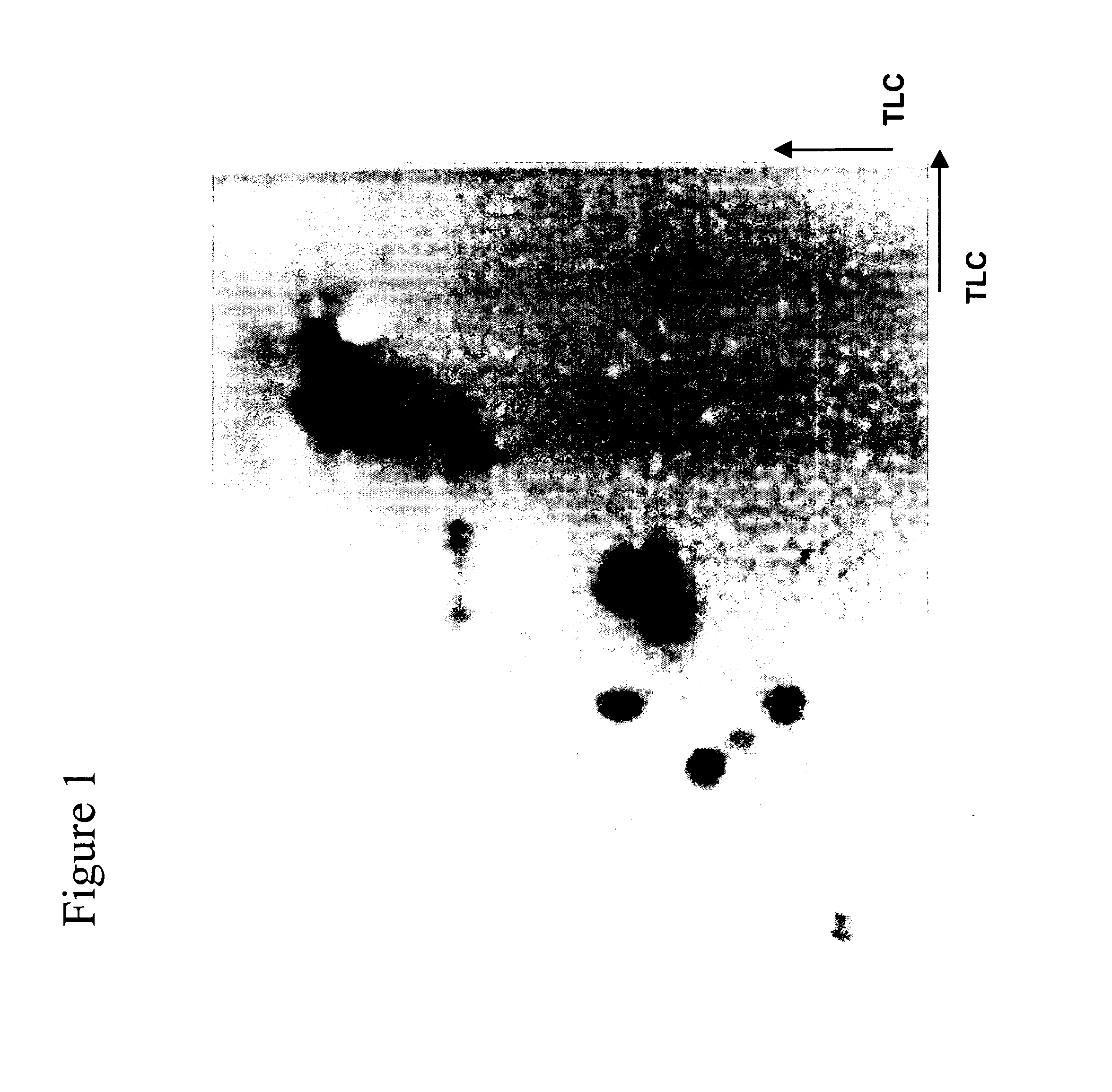
example 1
2D TLC Separation of Peptides
[0104] This example describes separation of peptides using TLC in two dimensions. Six model proteins were digested with trypsin and separated as described.
[0105] Trypsin (Sigma) was reconstituted in 1 mM HCl to obtain a concentration of 1 mg / mL. Model proteins β-casein and ovalbumin were dissolved in water and 10 mM ammonium carbonate, pH 8.5 buffer respectively. The final concentration in both cases was about 1 mg / mL. The proteins were denatured by heating them to 90° C. Subsequently, the trypsin and the protein were added together in a ratio of 1:10, according to the manufacturer recommended protocol and incubated for a day at 37° C. for 30 minutes. Formic acid (1%) was added to quench the reaction. Separation of the peptides obtained by tryptic digestion of proteins was performed using thin-layer chromatography (TLC) as follows: The separation was performed on HPTLC Cellulose plates or Silica gel 60 glass-backed HPTLC plates (Merck, Darmstadt, Germa...
example 2
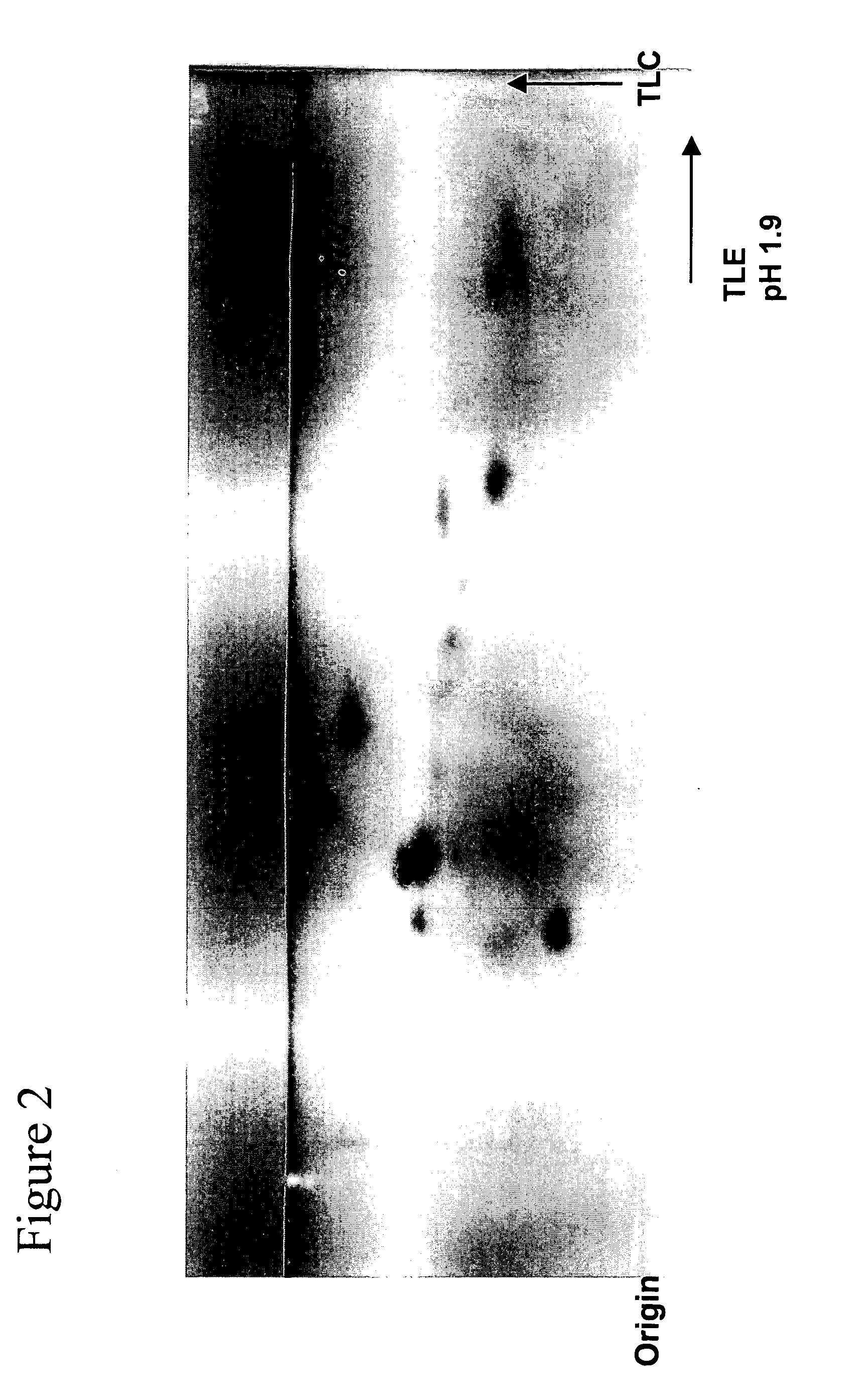
2D TLE / TLC Separation of Peptides
[0108] This example describes two dimension separation of peptides using thin-layer electrophoresis (TLE) and TLC. 2D TLE / TLC separation of peptide digests was accomplished by the well-established HTLE method. Briefly, 88% formic acid / glacial acetic acid / water, pH 1.9 (50:156:1794 v / v / v) was used as the mobile phase for the TLE dimension and n-butanol / pyridine / glacial acetic acid / water (75 / 50 / 15 / 60 v / v / v / v) was employed for the TLC dimension. TLE separations were performed using a Hunter Thin-Layer Electrophoresis (HTLE) instrument, Model HTLE-7002, CBS Scientific, Del Mar, Calif. Cellulose HPTLC plates were used for the separation.
[0109]FIG. 2 portrays a typical 2D separation of a O-casein tryptic peptide digest using a combination of TLE and TLC. Reasonable resolution separations of peptides are achieved by the TLE / TLC method. However, the separation results in a relatively low number of detected spots. Also, no anodically migrating peptides were...
example 3
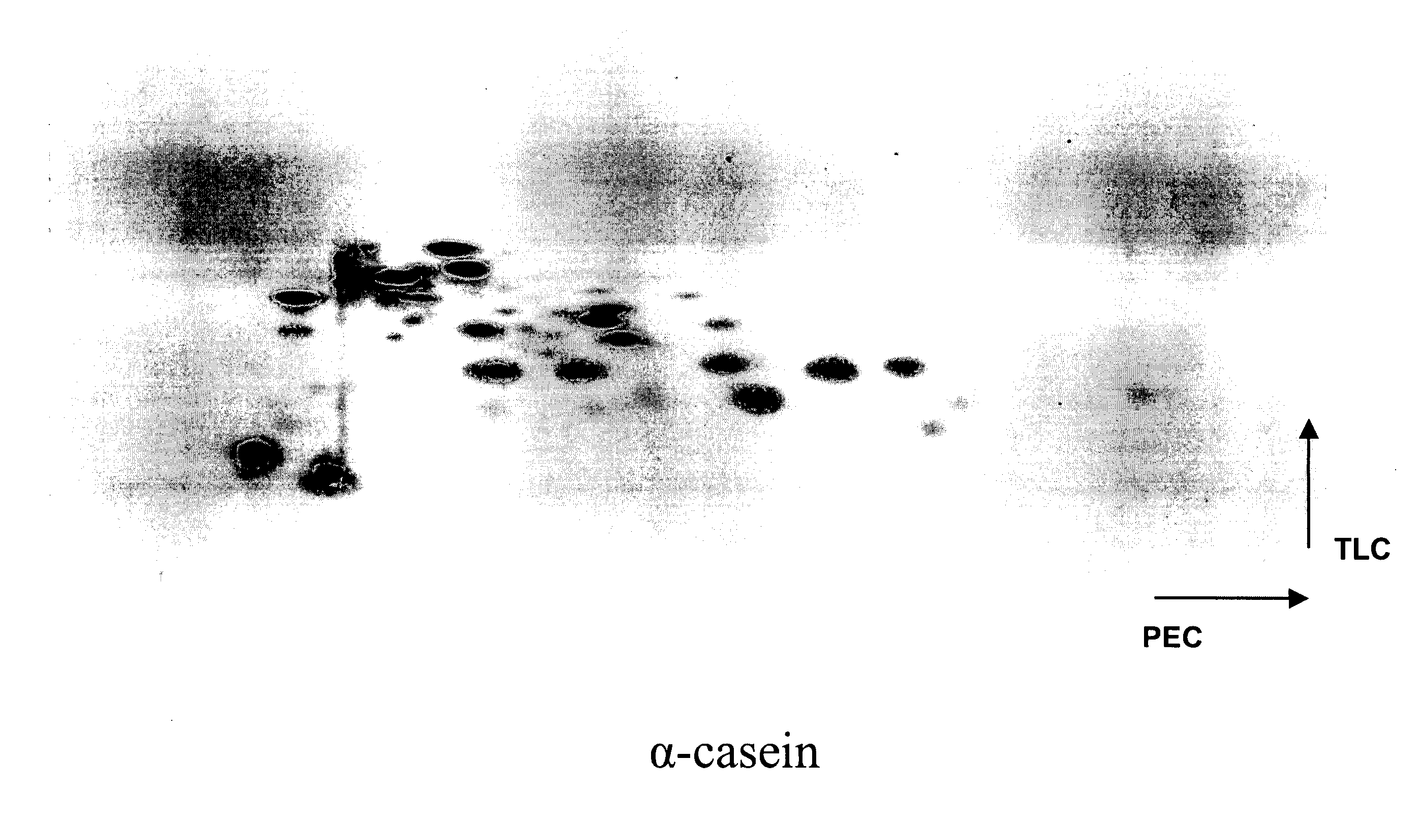
Two Dimensional (2D) PEC / TLC Separation of β-Casein and Ovalbumin Tryptic Peptide Digests
[0110] Tryptic peptide digests of ovalbumin and β-casein were performed as described in Example 1. Separations of tryptic digests of α-casein and ovalbumin were performed as described below.
[0111] The HTLE apparatus for PEC / TLE was assembled as per the manufacturer's instructions (CBS Scientific). Each of the buffer tanks was filled with approximately 500 mL of the buffer. Whatman 3 mM paper, folded in the middle, was used as a wick to transfer the solution from the buffer tanks (lined with platinum electrodes across the length of the tank) onto the separation plates. Wicks were made by cutting the paper to fit and overlap the TLC plate by about a cm from the edge of the plate. The wicks were then wetted in a glass tray, excess solvent drained by tapping on the sides of the tray and immersed in the buffer tanks on one edge leaving the other edge to overlap the plate. A pressure of about 0.7 At...
PUM
| Property | Measurement | Unit |
|---|---|---|
| Length | aaaaa | aaaaa |
| Force | aaaaa | aaaaa |
| Amphiphilic | aaaaa | aaaaa |
Abstract
Description
Claims
Application Information