Method For Preparing BioMedical Surfaces

a biomedical surface and surface technology, applied in the field of biomedical surfaces, can solve the problems of reducing the value of surfaces with recessed cavities, reducing the success rate of bone attachment, and often very expensive techniques to implement, and achieves less cost, stronger and robust interfacial adhesion, and enhanced bonding properties.

Active Publication Date: 2007-08-16
SYRACUSE UNIVERSITY
View PDF8 Cites 35 Cited by
  • Summary
  • Abstract
  • Description
  • Claims
  • Application Information

AI Technical Summary

Benefits of technology

[0008]It is an additional object and advantage of the present invention to provide a method for forming biomedical surfaces having nanoscale topography that provides faster, stronger and more robust interfacial adhesion to biological systems.
[0009]It is a further object and advantage of the present invention to provide a method for forming biomedical surfaces having nanoscale topography that is less costly.
[0010]It is a further object and advantage of the present invention to provide a method for forming biomedical surfaces having nanoscale topography that is applicable for use in connection with recessed cavities.
[0011]Other objects and advantages of the present invention will in part be obvious, and in part appear hereinafter.
[0012]In accordance with the foregoing objects and advantages, t

Problems solved by technology
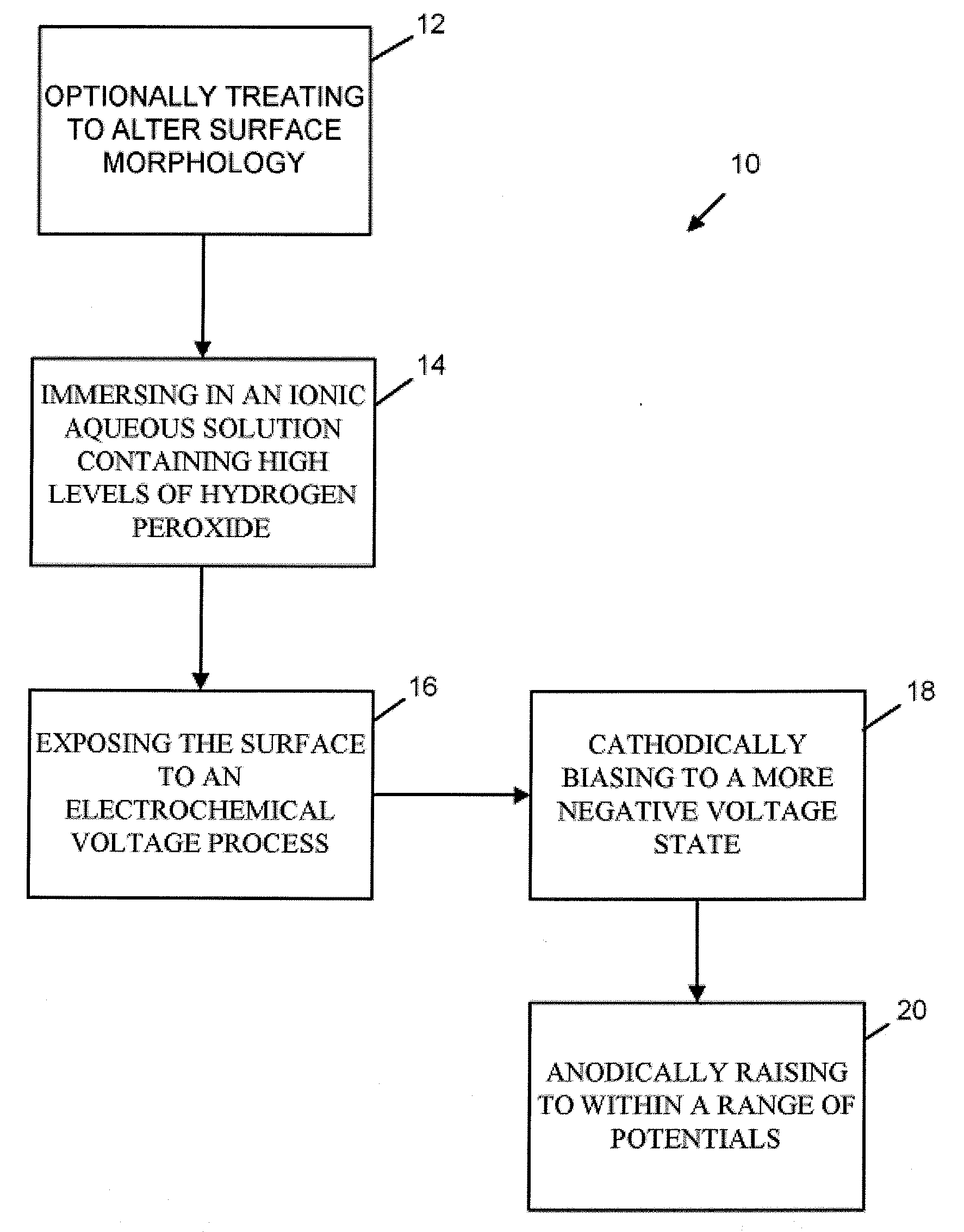
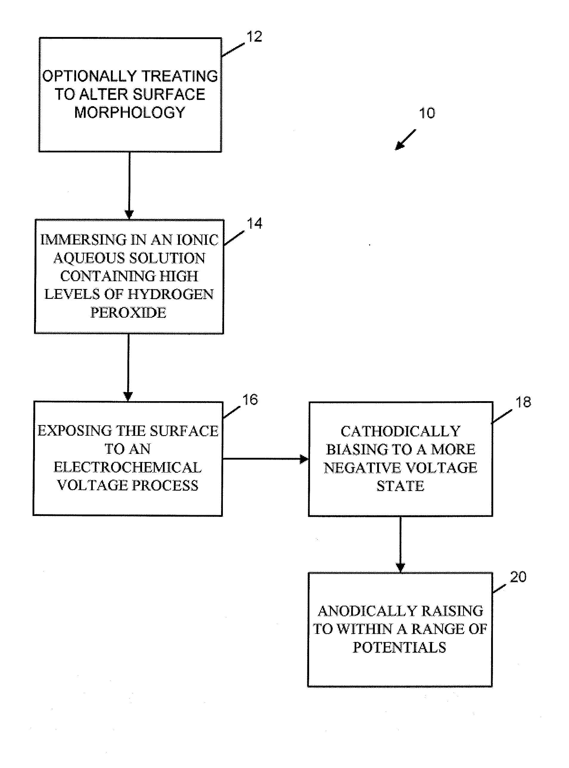
These techniques are frequently very expensive to implement and are of reduced value in

Method used


Image

  • Method For Preparing BioMedical Surfaces
  • Method For Preparing BioMedical Surfaces
  • Method For Preparing BioMedical Surfaces

Examples


example 1

[0030]Samples of medical grade Ti-6Al-4V-ELI (Extra-low-interstitial, ASTM F136 “Standard Specification for Wrought Titanium 6AL-4V ELI Alloy for Surgical Implant Applications”) were mechanically polished with increasing grit emory paper followed by polishing with 1.0, 0.3 and 0.05 μm Al2O3 particles suspended in water to yield a flat polished surface. These samples were then placed in an electrochemical cell and immersed in a PBS-H2O2 solution consisting of 0.154 M phosphate buffered saline (Sigma-Aldrich), and modified with 1 M H2O2. The sample was then electrochemical held at a voltage of −1V vs Ag / AgCl for 10 min, afterward the potential of the surface was scanned anodically at 1 mV / s up to 1 V vs Ag / Ag / Cl. At the end of this potential scan, the sample was removed from the solution, rinsed in DI water and allowed to dry. Afterward the sample was placed in the scanning electron microscope and imaged under secondary electrons. The resultant surface is seen in FIG. 5. The beta phas...

example 2

[0031]Samples of Ti-6Al-4V were prepared as in example 1 above. In this process, the sample is first held at a cathodic potential (−1V vs Ag / AgCl) for a period of 10 minutes and then immediately scanned up to more positive potentials at 1 mV / s and then held fixed for periods of time (typically 1000 s). The potential explored were: −0.5V, −0.1 V, and 0.1 V. These last two potentials relate to the active region of the polarization plots shown in FIG. 2, while −0.5V is still in the somewhat-cathodic potential region. The solution used to perform this was the same as that example 1 (1M H2O2 in PBS). After the process was complete, the samples were removed from the solution, rinsed in DI water, dried and imaged in the SEM. The results of Example 2 are seen in FIG. 6. The −0.5 V case (a) did not selectively dissolve the beta phase, while the other two (b) and (c) did.

example 3

[0032]The processed alloy was a commercially pure titanium alloy (ASTM F-36, grade 2). It was prepared and tested as described in example 1, where the potential was first held cathodic at −1.0 V and then scanned anodically to +1.0 V at 1 mV / s. Afterward, it was rinsed dried and imaged as above. The results are seen in FIG. 7, and indicate that the Cp-Ti surface developed micron-scale and nano-scale pits in the surface.

[0033]The method of the present invention may thus be used for the preparation of Ti-alloy surfaces for implantation into biological systems to enhance the interaction between the body and the implant. This process could be applied to a wide variety of medical devices where implant-tissue interactions are important. These include bone implant devices, dental implants and potential cardiovascular implants. The resulting effect allows modification of surfaces of titanium alloys to develop micron or smaller (nano-scale) surface topographic features that may enhance these ...


PUM

PropertyMeasurementUnit
Molar densityaaaaaaaaaa

Abstract

A method for selectively dissolving the beta (β) phase of a titanium alloy out of the surface of the alloy, thereby leaving behind a nano-scale porous surface having enhanced bonding properties with either a biological tissue, such as bone, or an adhesive material, such as a polymer or ceramic by immersing the alloy in an ionic aqueous solution containing high levels of hydrogen peroxide and then exposing the alloy to an electrochemical voltage process resulting in the selective dissolution of the beta phase to form a nano-topographic metallic surface.

Description

CROSS-REFERENCE TO RELATED APPLICATIONS[0001]This application claims priority to U.S. Provisional Patent Application Ser. No. 60 / 766,781, filed Feb. 10, 2006.BACKGROUND OF THE INVENTION[0002]1. Field of the Invention[0003]The present invention relates to biomedical surfaces and, more specifically, to a method of preparing biomedical surfaces for enhanced biological interaction and adhesion with adjoining materials or tissue.[0004]2. Description of the Related Art[0005]Porous surfaces have been used for over two decades to provide places for biological fixation between implant and tissue to take place. Typically these porous surfaces are processed by sintering, diffusion bonding, plasma spraying or other mechanism and create rough bead-like or fiber-like surfaces. The spaces between these materials then serve as sites where bone or other tissue can in-grow and attach to the implant. This process is called biological fixation and is used extensively in implants (e.g., dental, hips and...

Claims


Application Information

Patent Timeline
IPC IPC(8): C25D5/00H01L21/00
CPCA61L27/06A61L27/50A61L2400/18Y10T428/24355A61F2310/00023A61F2002/3084A61F2/30771C25D11/26C25F3/08
Inventor GILBERT, JEREMYBAI, ZHIJUNCHANDRASEKARAN, NITHYA
Owner SYRACUSE UNIVERSITY
Why Eureka
Try Eureka