
Method for detecting an inflammatory disease or cancer

Inactive Publication Date: 2008-01-24
FRANTZ BIOMARKERS
View PDF9 Cites 13 Cited by
  • Summary
  • Abstract
  • Description
  • Claims
  • Application Information

AI Technical Summary

Benefits of technology

[0041] It is a still further object of the present invention to provide a non-invasive method to detect the occurrence of ovulation during a menstrual cycle.
[0042] It is another object of the present invention to provide a non-invasive method to monitor the presence of an inflammatory disease or a cancer over time.

Problems solved by technology

Many of these diseases are debilitating and are becoming increasingly common in our society.
Malfunctions in the repair of this oxidative damage can lead to ovarian cancer, a condition that causes further inflammation and oxidative stress.
Ovarian cancer is one of the deadliest cancers for women, due to its high fatality rate.
Although CA-125 is elevated in 82% of women with advanced ovarian cancer, it has very limited clinical application for the detection of early stage disease, exhibiting a positive predictive value of less than 10%.

Method used


Image

  • Method for detecting an inflammatory disease or cancer
  • Method for detecting an inflammatory disease or cancer
  • Method for detecting an inflammatory disease or cancer

Examples


example 1

Plasmenyl-PE in Serum Samples

[0167] Materials

[0168] 18:0, 22:6 PPE; 18:0, 20:4 PPE; and 18:0, 18:1 PPE were purchased from Avanti Polar Lipids (Alabaster, Ala., USA). Using these lipids, it was determined that the MRM transition of 18:0, 22:6 PPE; 18:0, 20:4 PPE; 18:0, 18:1 PPE; 18:0, 18:2 PPE; 16:0 22:6 PPE; 16:0, 20:4 PPE; 16:0, 18:1 PPE; and 16:0, 18:2 PPE were 774.2→327.2, 750.2→303.2, 728.2→281.2, 726.2→279.2, 746.2→327.2, 722.2→303.2, 700.2→281.2, and 698.2→279.2 respectively.

Example 1(a)

Extraction of Plasmenyl-PE from Serum Samples

[0169] Lipid extraction was done according to the following procedure: Add 50 μL 10 μM 1,2-diheptadecanoyl-sn-glycerol-3-phosphoethanolamine, the internal standard for the assay, into 50 μl serum samples. Vortex and add 2 ml 2:1 methanol-chloroform into the samples. Vortex again and centrifuge the mixture for 5 minutes at 4000 rpm and 10° C. Transfer the upper liquid layer into a test tube and dry the liquid layer under nitrogen. Then add 400 μ...

example 1 (

Example 1(c)

Samples and Statistical Analysis

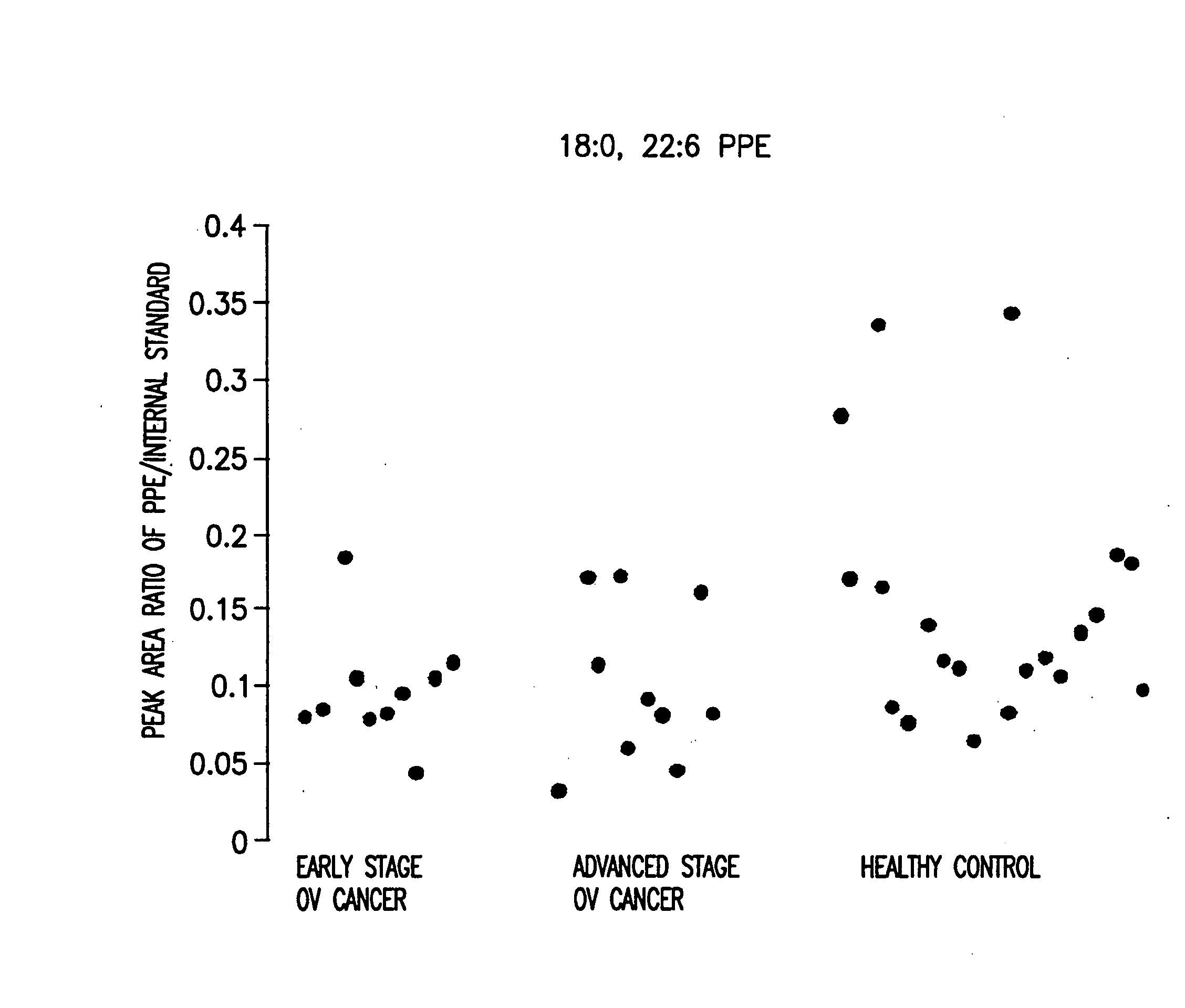
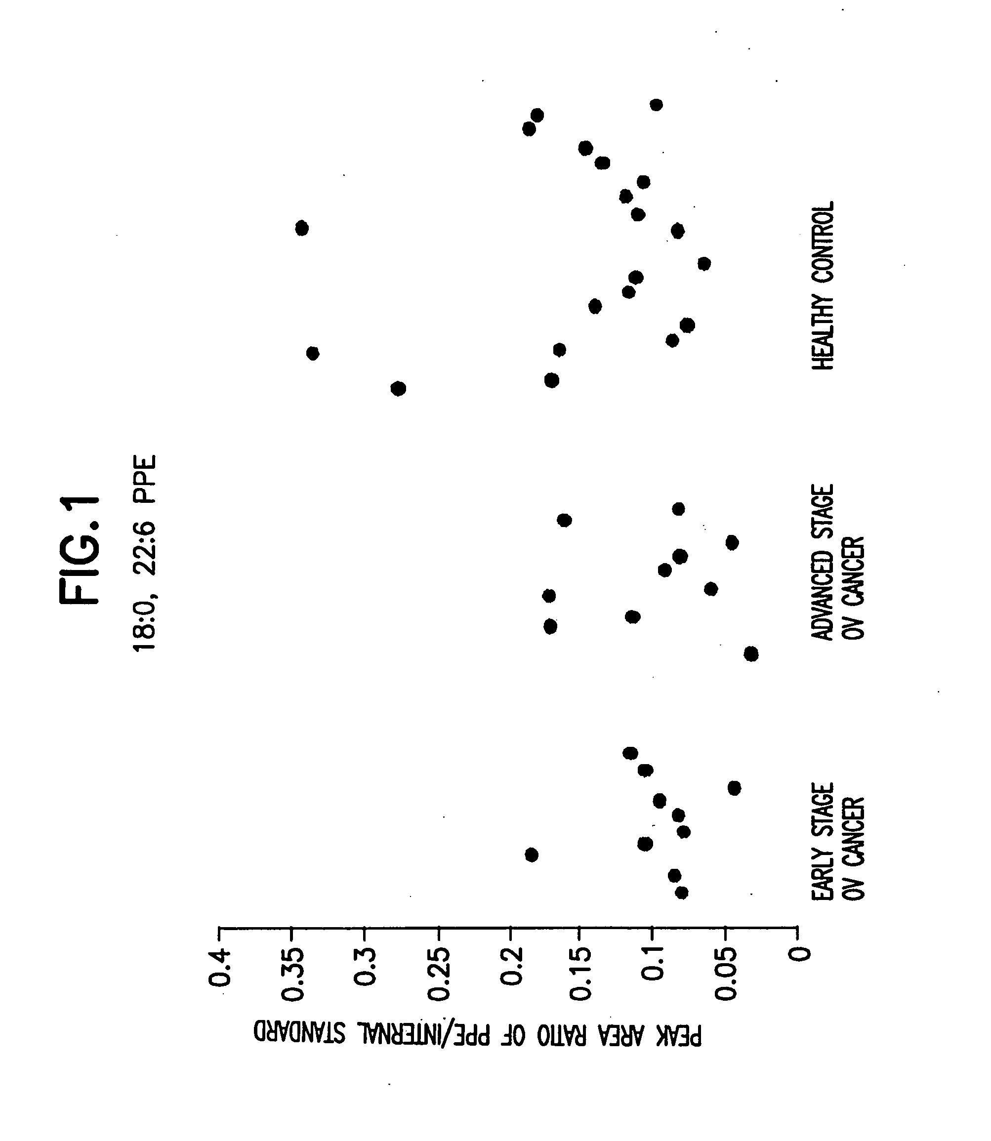
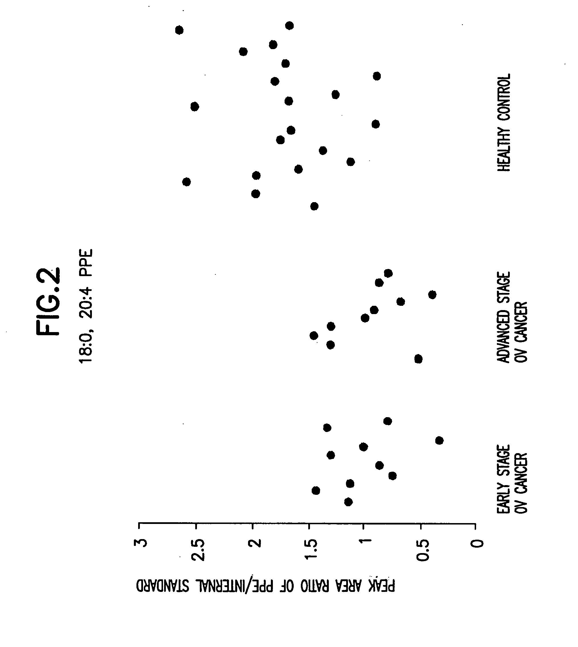
[0171] 40 serum samples were collected. Among them were 10 early stage ovarian cancer, 10 late stage ovarian cancer, and 20 healthy control. Data analysis was done using the student t-test and the peak area ratio of analyte to internal standard was determined. The results are shown in Table 1 and FIG. 1 to FIG. 8.

TABLE 1Level of 18:0, 18:2 plasmenyl-PE (see FIG. 4), standard deviation,and p value (related to healthy control samples) in 40 serum samples,as determined by peak ratio of analyte to internal standardLevel of 18:0, 18:2StandardSerum sampleplasmenyl-PEDeviationp valueEarly stage ovarian cancer0.5370.322Advanced stage ovarian0.5550.397cancerHealthy control1.110.298—

[0172] If 0.70 is used as the cut-off, the levels of 18:0, 18:2 plasmenyl-PE in 8 of 10 early stage ovarian cancer patients are below this value, with the sensitivity equaling 80%. The levels of 18:0, 18:2 plasmenyl-PE in-8 of 10 advanced stage ovarian cancer are belo...

example 2

Plasmenyl-PE in Plasma Samples

[0173] Materials

[0174] 18:0, 22:6 PPE; 18:0, 20:4 PPE; 18:0, 18:1 PPE were purchased from Avanti Polar Lipids (Alabaster, Ala., USA). Using these lipids, it was determined that the MRM transition of 18:0, 22:6 PPE; 18:0, 20:4 PPE; 18:0, 18:1 PPE; 18:0, 18:2 PPE; 16:0 22:6 PPE; 16:0, 20:4 PPE; 16:0, 18:1 PPE; and 16:0, 18:2 PPE were 774.2→3.27.2, 750.2→303.2, 728.2→281.2, 726.2→279.2, 746.2→327.2, 722.2→303.2, 700.2→281.2, 698.2→279.2 respectively.

Example 2(a)

Extraction of Plasmenyl-PE from Plasma Samples

[0175] Lipid extraction was done according to the following procedure: Add 200 μL 10 μM 1,2-diheptadecanoyl-sn-glycerol-3-phosphoethanolamine, the internal standard for the assay, into 50 μl plasma samples. Vortex and add 2 ml 2:1 methanol-chloroform into the samples. Vortex again and centrifuge the mixture for 5 minutes at 4000 rpm and 10° C. Transfer the upper liquid layer into a test tube and dry the liquid layer under nitrogen. Then add 400 μl 0...


PUM


Abstract

A method of detecting an inflammatory disease or a cancer in a test subject comprising determining the amount of plasmenyl-PE or a biomarker having a mass charge ratio of approximately 698.2, 722.2, 726.2 or 750.2 in a sample of bodily fluid taken from the test subject and comparing the amount of plasmenyl-PE (or the biomarker) in the sample of the bodily fluid from the test subject to a range of amounts of plasmenyl-PE (or the biomarker) found in samples of the bodily fluid from a group of normal subjects of the same species as the test subject and lacking the inflammatory disease or the cancer, whereby a change in the amount of the plasmenyl-PE (or the biomarker) (such as a lower amount) in the sample of the bodily fluid from the test subject indicates the presence of the inflammatory disease or the cancer.

Description

CROSS-REFERENCE TO RELATED APPLICATIONS [0001] This application claims the benefit under 35 USC 119(e) of provisional application Ser. No. 60 / 738,849 filed Nov. 22, 2005 and provisional application Ser. No. 60 / 843,088 filed Sep. 8, 2006, the entire contents of both of which provisional applications are incorporated by reference herein.BACKGROUND OF THE INVENTION [0002] 1. Field of the Invention [0003] A method of detecting an inflammatory disease or cancer in a test subject. The present invention is further directed to a method for detecting the occurrence of ovulation during a menstrual cycle. More particularly, the present invention relates to a method for detecting inflammatory disease or cancer in a test subject by determining the amount of a plasmalogen, such as plasmenyl-PE (“PPE”), in a sample of bodily fluid taken from the test subject. The present invention is particularly useful as a screening test for cancer, such as ovarian cancer. [0004] 2. Background Information [0005]...

Claims


Application Information

Patent Timeline
IPC IPC(8): G01N33/92
CPCG01N33/57449G01N2800/361G01N33/92G01N33/6893
Inventor SHAN, LIANDAVIS, LORELEI D.
Owner FRANTZ BIOMARKERS
Learn More