Nanoparticle Mediated Ultrasound Therapy and Diagnostic Imaging

a nanoparticle and ultrasound technology, applied in the field of nanoparticle mediated ultrasound therapy and diagnostic imaging, can solve the problems of low power density ultrasound not being useful for treating cells or tissues, damage to adjacent healthy cells or tissues, combined use of ultrasound and targeted contrast agents has a basic limitation, etc., to achieve precise imaging of diseased tissue borders, enhance the effect of hyperthermia, and localization

Inactive Publication Date: 2008-02-21
KISLEV HANOCH
View PDF24 Cites 230 Cited by
  • Summary
  • Abstract
  • Description
  • Claims
  • Application Information

AI Technical Summary

Benefits of technology

[0026] The present invention provides systems and methods for use in cell and tissue therapy and imaging. The primary object of the present invention is to provide a system and method for inducing enhanced, localized, targeted hyperthermia in such cell and tissue, while minimizing collateral damage to surrounding normal cells and tissue, as well as providing a means for precise imaging of the diseased tissue borders and volume.

Problems solved by technology

Low power density ultrasound is not useful for treating cells or tissues by hyperthermia due to only a slight difference between the absorption rate of a diseased and a healthy tissue and the acoustic properties variations in the human body, which may cause damage to adjacent healthy cells or tissues.
However, the combined use of ultrasound and targeted contrast agents has a basic limitation: it is very hard to accumulate the necessary concentration of contrast agent within the targeted tissue volume.
An attempt to load the desired contrast agent concentration often results in forming a microbubble layer around the targeted tissue, which may block the ultrasound radiation from penetrating into the targeted tissue volume.
When provided through a coupling device, ultrasound passing through intervening tissues is usually of low intensity and therefore, relatively non-destructive.
However, at the focal point, the accumulated energy is raised to a pre-determined higher intensity and tissue destruction occurs at, or around, the focal point.
However, in many cases the focused ultrasound energy generates a dense microbubble cloud between the targeted and healthy tissue, which blocks the ultrasound radiation.
In addition, dense microbubble cloud interaction with HIFU involves the potential occurrence of cavitation events, which, in turn, leads to the formation of destructive, or possibly mutagenic, free radicals [Miller et al., Ultrasound in Med.
Furthermore, the complex nature of the human organs complicates the aiming procedure when using HIFU, and requires the use of guided treatment, typically using on-line magnetic resonance imaging (MRI) instrumentation.
The volume and speed of HIFU treatments is limited by the potential destruction of normal tissue within the near field between the target and the ultrasound probe and due to targeting errors.
However, the use of NIR absorbing nanoparticles for photothermal therapy suffers from a basic drawback: the vast nanoparticle volume concentration required for localized heating.
The extremely slow diffusion rate of NIR optimized nanoshells (with a diameter of about 200 nm) makes it very hard to accumulate such concentrations within the volume of a tumor whose size is larger than one cm.
Another unsolved problem of photothermal treatment is the total penetration depth of electromagnetic radiation including NIR, in a tissue.
Exposure above threshold may induce deep burns, partially due to sub-dermal back scattering which increases the local internal tissue damage near the light source.
Unfortunately, the size of nanoparticles which can penetrate through malignant blood vessels (40-200 nm) relates to a very small interaction cross section at the linear range of interaction with ultrasound radiation.
However, the required nanoparticle concentration was found to be 1*1011 particles / cm3, which concentration is extremely hard to achieve in the tumor volume.
Unfortunately, there are minor differences between the acoustic impedance difference of healthy and diseased tissue.
Unfortunately, nanoparticles whose size is smaller than 100 nm may carry negligible gas content which is hardly practical for imaging purposes.

Method used


Image

  • Nanoparticle Mediated Ultrasound Therapy and Diagnostic Imaging
  • Nanoparticle Mediated Ultrasound Therapy and Diagnostic Imaging
  • Nanoparticle Mediated Ultrasound Therapy and Diagnostic Imaging

Examples


Embodiment Construction

Definitions

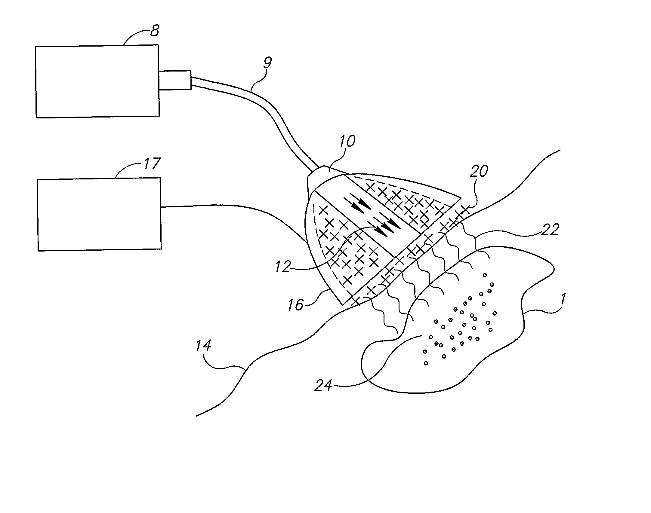
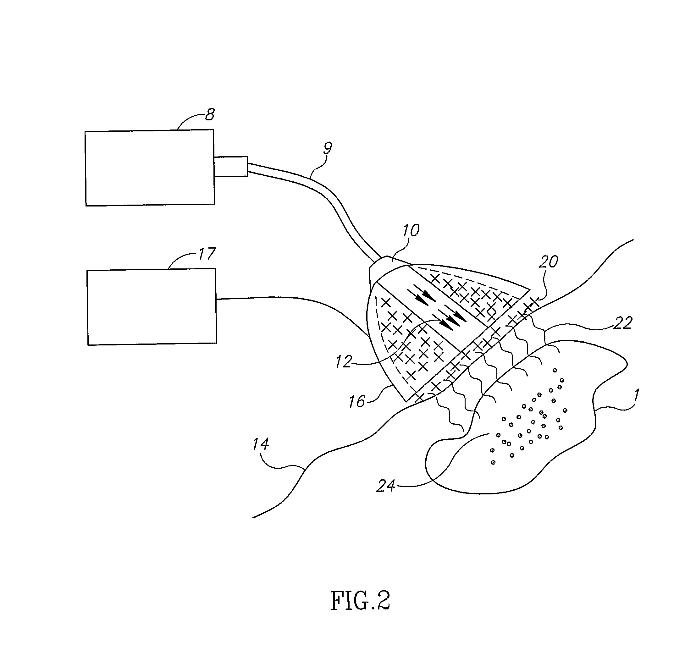
[0085] As used herein, “energy source” encompasses any and all forms of excitation, including radiation from any or all regions of the electromagnetic spectrum, ultrasound, magnetic fields, electric fields, microwave radiation, laser radiation, etc.

[0086] As used herein, “light” means electromagnetic radiation.

[0087] As used herein, “electromagnetic radiation” is defined as radiation having an electric field and a magnetic field propagating at right angles to one another and is further limited to only the following: microwaves, infrared, visible, ultraviolet, x-rays, gamma rays, and cosmic rays. As used herein, “electromagnetic radiation” does not include radio-frequency radiation.

[0088] As used herein, “nanoparticle” is defined as a particle having a diameter of from 1 to 1000 nanometers, having any size, shape, structure or morphology exhibiting enhanced absorption of electromagnetic radiation in a relatively narrow spectral band, between 300 nm and 2000 nm, as a si...


PUM


Abstract

The present invention relates to systems and methods for localized delivery of heat, useful for localized imaging and treatment of a biological material. The systems and methods of the invention can be utilized for localized treatment of cancer, inflammation or other disorders involving over proliferation of tissue, and for tissue repair. The method comprises exposing nanoparticles to electromagnetic radiation under conditions wherein the nanoparticles generate microbubbles which emit heat when exposed to ultrasonic radiation.

Description

FIELD OF THE INVENTION [0001] The present invention relates to a system and method for localized delivery of heat, useful for localized imaging and treatment of a biological material, particularly for tissue repair and localized treatment of cancer, inflammation or other disorders involving over proliferation of tissue. The methods comprise exposing nanoparticles to electromagnetic radiation under conditions wherein the nanoparticles generate microbubbles which emit and propagate heat when further exposed to ultrasonic radiation. BACKGROUND OF THE INVENTION [0002] Localized heating of cells and tissues is desirable in many applications. Precise, localized heating has been shown to have therapeutic benefits, while minimizing the collateral damage to nearby cells and tissue. The therapeutic effects of thermal ablation range from the destruction of cancerous cells and tumors, to the therapeutic or cosmetic removal of benign tumors and other undesirable tissues. [0003] In addition to th...

Claims


Application Information

Patent Timeline
Patent Type & Authority Applications(United States)
IPC IPC(8): A61N7/02A61N5/00
CPCA61B5/0059A61B5/411A61B5/415A61B5/418A61N2007/0039A61B18/28A61N7/00A61N7/022A61B8/481A61B8/4444
Inventor KISLEV, HANOCH
Owner KISLEV HANOCH
Why Eureka
Try Eureka