

Novel Dyes for Organ Function Monitoring
a technology of optical probes and organ function, applied in the field of new optical probes for organ function monitoring, can solve the problems of inability to correlate single value returned several hours after sampling with other important physiologic events, inability to obtain new or repeat data, and inability to accurately predict the effect of organ function, etc., to achieve simple, efficient and effective monitoring of organ function.
- Summary
- Abstract
- Description
- Claims
- Application Information
AI Technical Summary
Benefits of technology
Problems solved by technology
Method used
Image

Examples
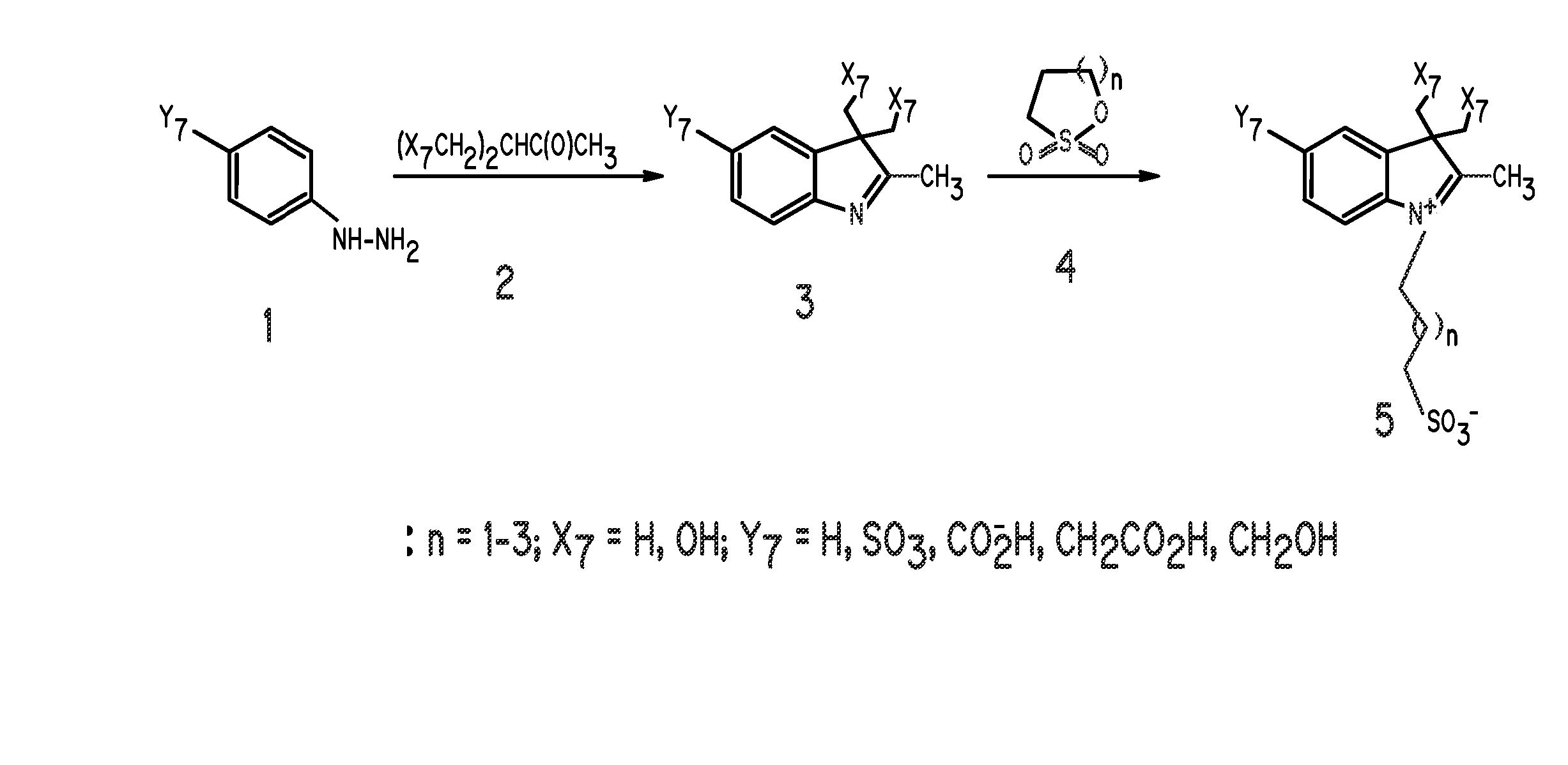
example 1
Synthesis of Indole Disulfonate
FIG. 1, Compound 5, Y7═SO3−; X7═H; n=1
[0066] A mixture of 3-methyl-2-butanone (25.2 mL), and p-hydrazinobenzenesulfonic acid (15 g) in acetic acid (45 mL) was heated at 110° C. for 3 hours. After reaction, the mixture was allowed to cool to room temperature and ethyl acetate (100 mL) was added to precipitate the product, which was filtered and washed with ethyl acetate (100 mL). The intermediate compound, 2,3,3-trimethylindolenium-5-sulfonate (FIG. 1, compound 3) was obtained as a pink powder in 80% yield. A portion of compound 3 (9.2 g) in methanol (115 mL) was carefully added to a solution of KOH in isopropanol (100 mL). A yellow potassium salt of the sulfonate was obtained in 85% yield after vacuum-drying for 12 hours. A portion of the 2,3,3-trimethylindolenium-5-sulfonate potassium salt (4 g) and 1,3-propanesultone (2.1 g) was heated in dichlorobenzene (40 mL) at 110° C. for 12 hours. The mixture was allowed to cool to room temperature and the re...
example 2
Synthesis of Indole Disulfonate
FIG. 1, Compound 5, Y7═SO3−; X7═H; n=2
[0068] This compound was prepared by the same procedure described in Example 1, except that 1,4-butanesultone was used in place of 1,3-propanesultone.
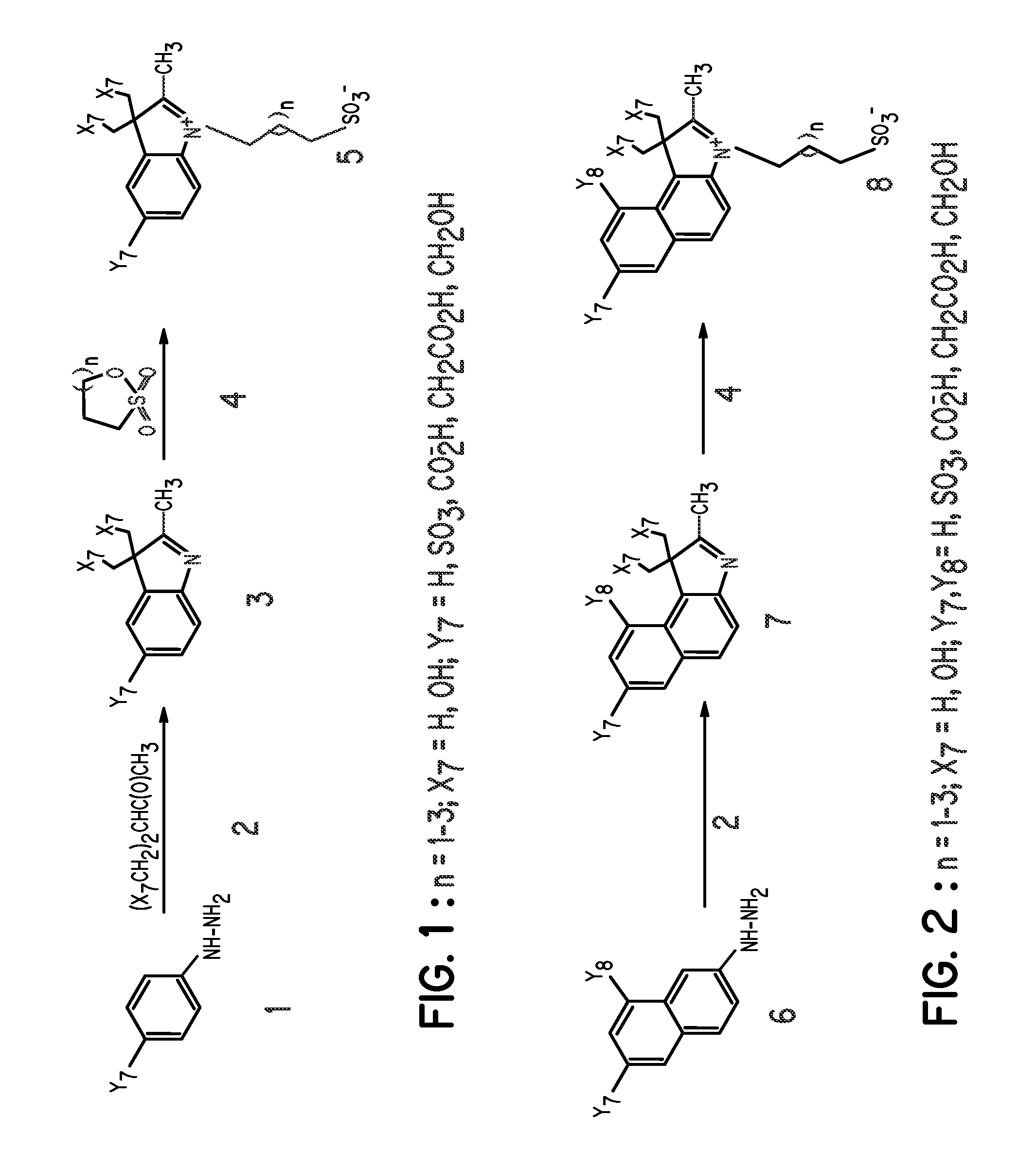
example 3
Synthesis of Benzoindole Disulfonate
FIG. 2, Compound 8, Y7, Y8═SO3−; X7═H; n=2
[0069] This compound was prepared by the same procedure described in Example 1, except that hydrazinonaphthalenedisulfonic acid was used in place of hydrazinobenzenesulfonic acid.
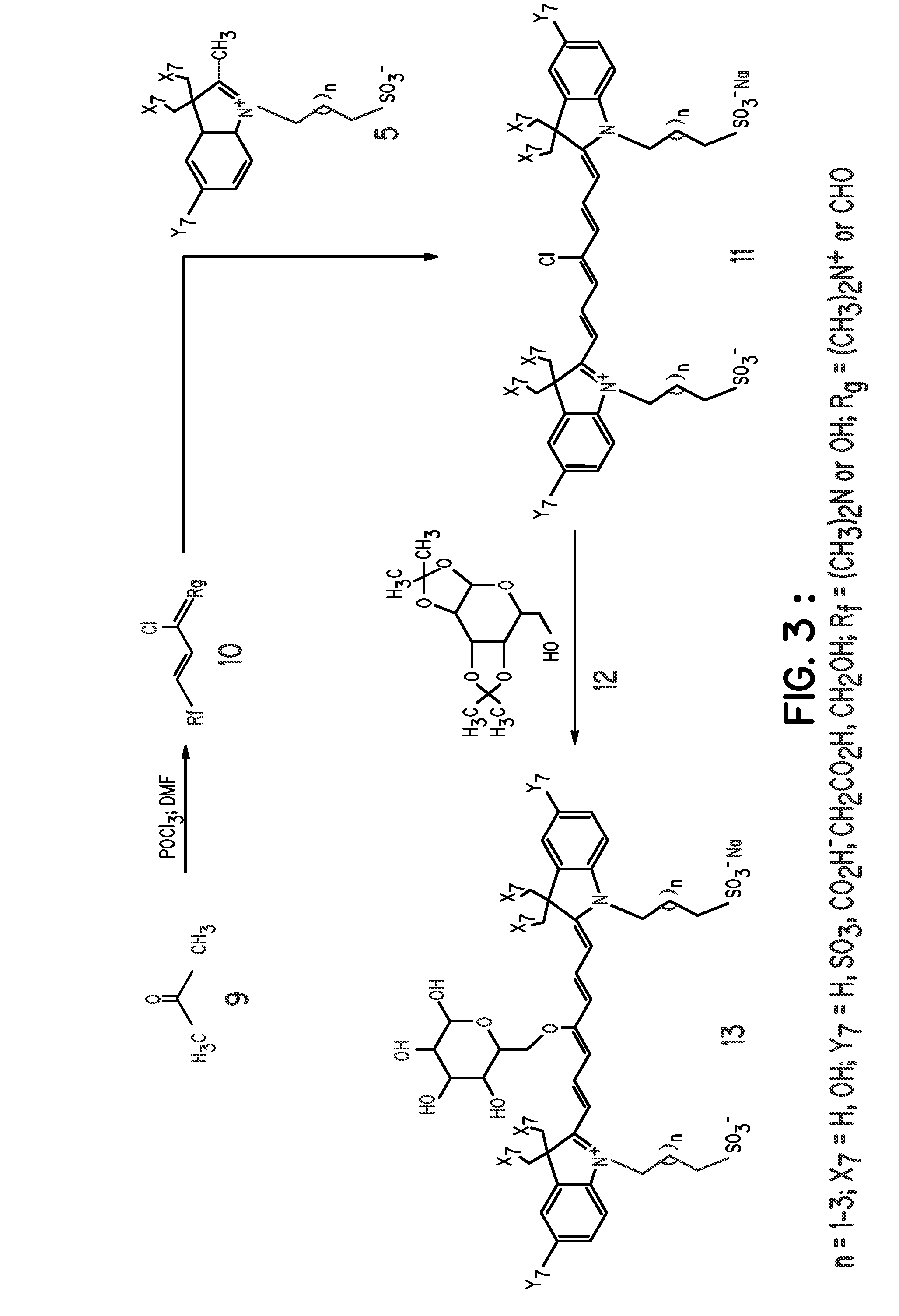
[0070] Other compounds prepared by a similar method include polyhydroxyindoles such as:
PUM
| Property | Measurement | Unit |
|---|---|---|
| of wavelength | aaaaa | aaaaa |
| light of wavelength | aaaaa | aaaaa |
| wavelength | aaaaa | aaaaa |
Abstract
Description
Claims
Application Information