Methods of In Vitro Analysis Using Time-Domain Nmr Spectroscopy

a time-domain, nmr spectroscopy technology, applied in the direction of analysis using nuclear magnetic resonance, using reradiation, instruments, etc., can solve the problems of high cost of nmr systems that utilize superconducting magnets, cryogenic cooling, and limited field strength

Active Publication Date: 2008-04-17
ASCENSIA DIABETES CARE HLDG AG
View PDF12 Cites 5 Cited by
  • Summary
  • Abstract
  • Description
  • Claims
  • Application Information

AI Technical Summary

Benefits of technology

[0009] According to one method, an analyte concentration of a sample is determined in vitro by placing the sample into a low-field, bench-top time-domain nuclear magnetic resonance (TD-NMR) spectrometer. The NMR spectrometer is tuned to measure a selected type of atom. A magnetic field is applied to the sample using a fixed, permanent magnet. At least one 90 degree radio-frequency pulse is applied to the sample. The 90 degree radio-frequency

Problems solved by technology

Although permanent magnets provide acceptable field stability and are less costly, the field strength is limited to approximately 1.4 T (60 MHz).
Unfortunately, NMR systems that utilize superconducting magnets are very expensive, require

Method used


Image

  • Methods of In Vitro Analysis Using Time-Domain Nmr Spectroscopy
  • Methods of In Vitro Analysis Using Time-Domain Nmr Spectroscopy
  • Methods of In Vitro Analysis Using Time-Domain Nmr Spectroscopy

Examples


Embodiment Construction

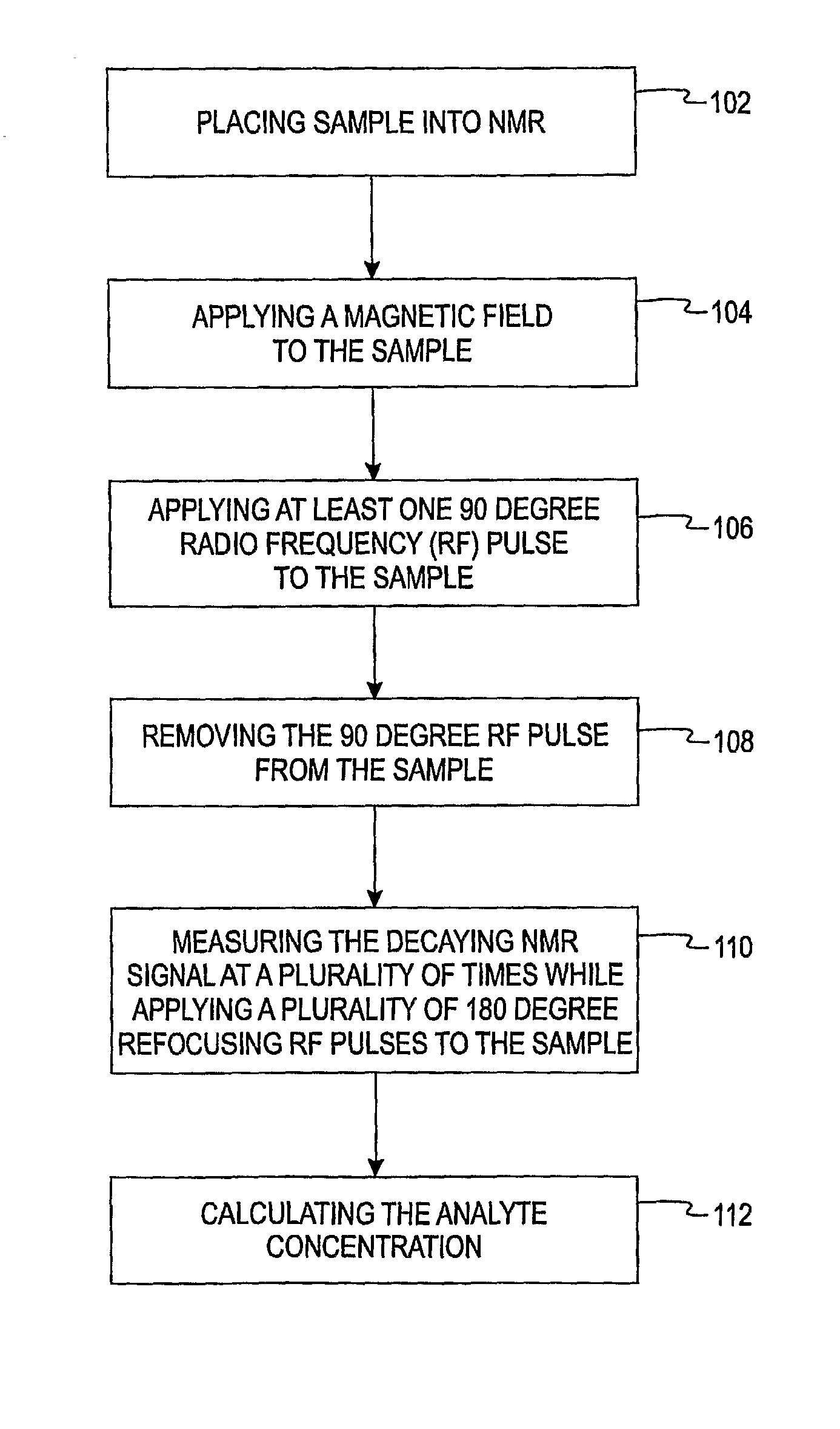
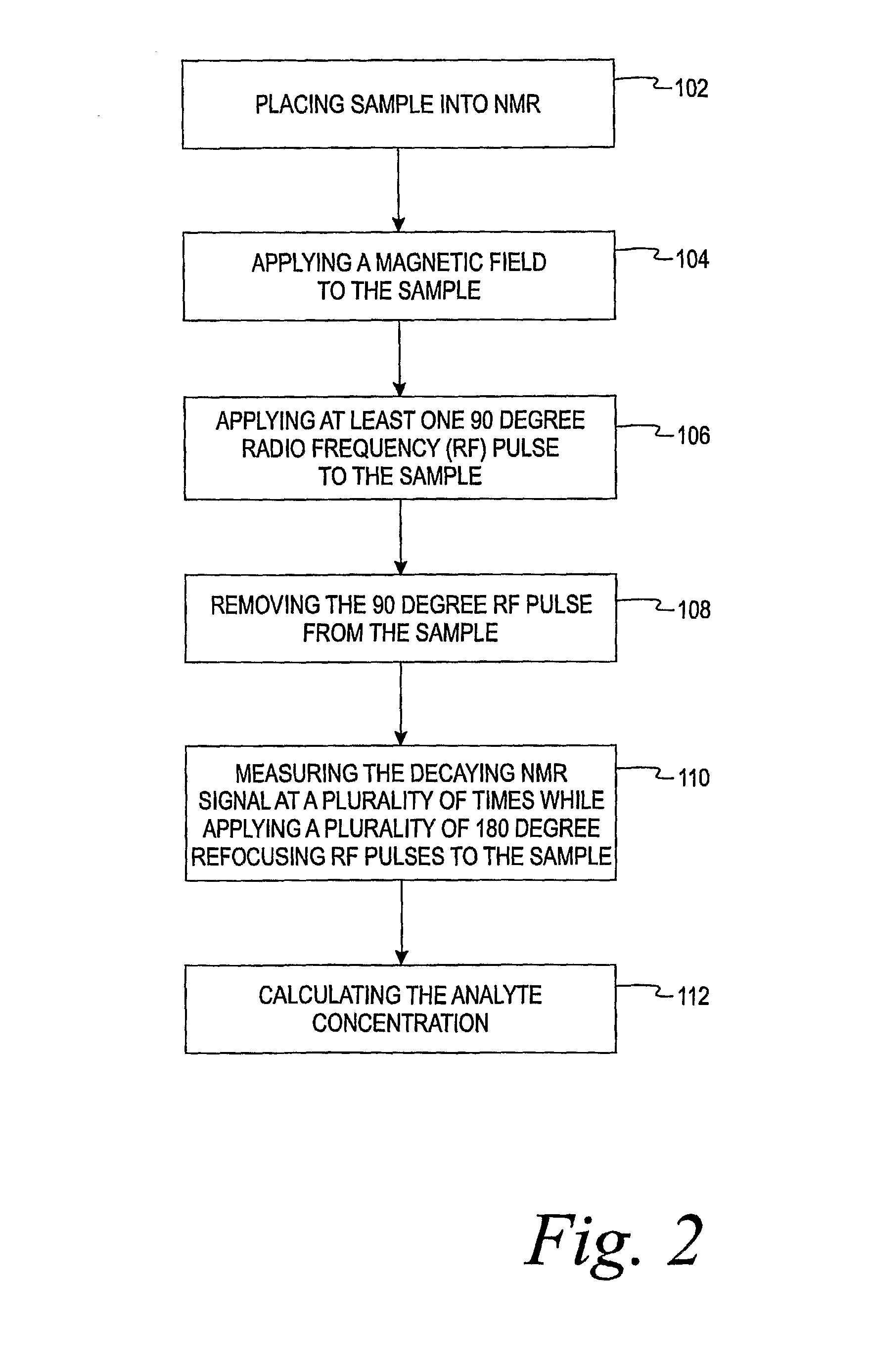
[0012] The present invention is directed to a method of using a time-domain nuclear magnetic resonance (TD-NMR) spectrometer to determine the analyte concentration of a sample. The method is done in vitro and may be performed without using reagents. The term, in vitro, is defined herein as being an artificial environment outside the living organism (e.g., a petri dish or test tube). The present invention is anticipated to be used in hospitals and clinics, but it is contemplated that it may be used in other locations.

[0013] Some non-limiting examples of analytes that may be monitored in certain individuals include glucose, cholesterol (e.g. HDL, LDL, total), triglycerides, globulin, albumin, total protein, blood urea nitrogen, alkaline phosphastase, and creatinine. The present invention is not limited, however, to these specific analytes. The analytes may be in, for example, body fluid such as a blood serum sample, a blood plasma sample or a urine sample.

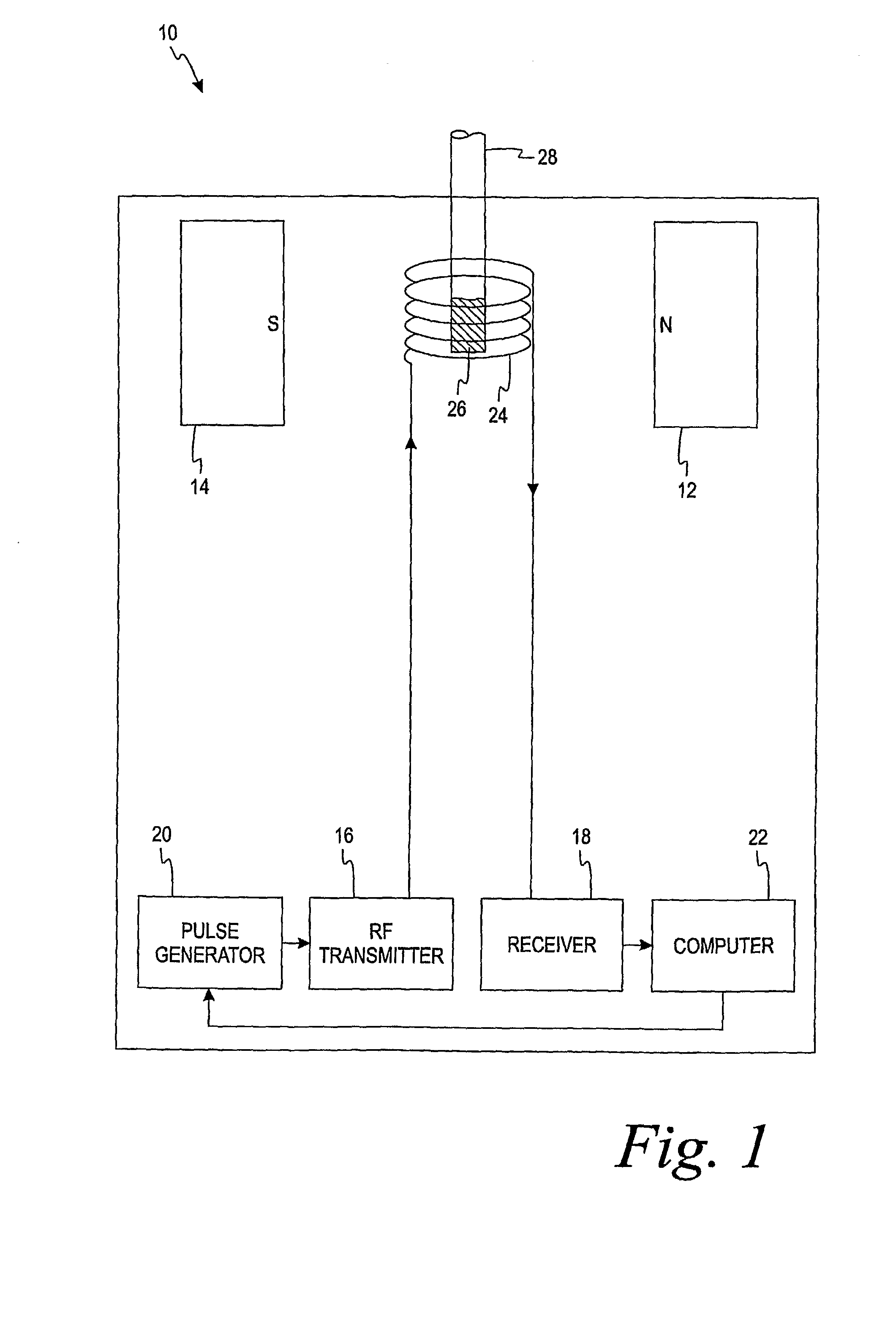
[0014] The spectrometer is ...


PUM


Abstract

An in vitro method of determining an analyte concentration of a sample includes placing the sample into a low-field, bench-top time-domain nuclear magnetic resonance (TD-NMR) spectrometer. The NMR spectrometer is tuned to measure a selected type of atom. A magnetic field is applied to the sample using a fixed, permanent magnet. At least one 90 degree radio-frequency pulse is applied to the sample. The radio-frequency pulse is generally perpendicular to the magnetic field. The 90 degree radio-frequency pulse is removed from the sample so as to produce a decaying NMR signal. The decaying NMR signal is measured at a plurality of times while applying a plurality of 180 degree refocusing radio-frequency pulses to the sample. The analyte concentration is calculated from the plurality of measurements associated with the decaying NMR signal and a selected model.

Description

FIELD OF THE INVENTION [0001] The present invention relates generally to methods of determining an analyte concentration of a sample. More particularly, the present invention relates to methods of determining an analyte using a low-field, time-domain nuclear magnetic resonance (NMR) spectrometer. BACKGROUND OF THE INVENTION [0002] The quantitative determination of analytes in body fluids is of great importance in the diagnoses and maintenance of certain physiological abnormalities. Some non-limiting examples of analytes that may be monitored in certain individuals include cholesterol (e.g. HDL, LDL, total), triglycerides, globulin, albumin, total protein, blood urea nitrogen, creatinine, and alkaline phosphastase. [0003] Nuclear magnetic resonance (NMR) spectroscopy is an analytical and diagnostic technique that is used for structural and quantitative analysis of one or more analytes in a sample. One specific type of NMR is time domain nuclear magnetic resonance (TD-NMR). TD-NMR spe...

Claims


Application Information

Patent Timeline
IPC IPC(8): G01R33/465C12Q1/42
CPCG01R33/465G01N24/08
Inventor CARPENTER, SCOTT E.BENSON, THOMAS P.
Owner ASCENSIA DIABETES CARE HLDG AG
Why Eureka
Try Eureka